 Movie
Movie Controller

[English] 日本語
 Yorodumi
- PDB-4c7w: Crystal structure of Mouse Hepatitis virus strain S Hemagglutinin... -
+ Open data

- Basic information
| Entry | Database: PDB / ID: 4c7w | |||||||||
|---|---|---|---|---|---|---|---|---|---|---|
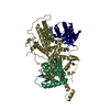
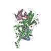
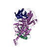
| Title | Crystal structure of Mouse Hepatitis virus strain S Hemagglutinin- esterase in complex with 4-O-acetylated sialic acid | |||||||||
 Components Components | HEMAGGLUTININ-ESTERASE | |||||||||
 Keywords Keywords | HYDROLASE / RECEPTOR DESTROYING / RECEPTOR BINDING | |||||||||
| Function / homology |  Function and homology information Function and homology informationsialate 9-O-acetylesterase activity / sialate 4-O-acetylesterase activity / sialate O-acetylesterase / host cell surface receptor binding / fusion of virus membrane with host plasma membrane / viral envelope / host cell plasma membrane / virion membrane / membrane Similarity search - Function | |||||||||
| Biological species |  MURINE HEPATITIS VIRUS MURINE HEPATITIS VIRUS | |||||||||
| Method |  X-RAY DIFFRACTION / SYNCHROTRON / MOLECULAR REPLACEMENT / Resolution: 2.5 Å X-RAY DIFFRACTION / SYNCHROTRON / MOLECULAR REPLACEMENT / Resolution: 2.5 Å | |||||||||
 Authors Authors | Zeng, Q.H. / Huizinga, E.G. | |||||||||
 Citation Citation | Journal: Plos Pathog. / Year: 2012 Title: The Murine Coronavirus Hemagglutinin-Esterase Receptor-Binding Site: A Major Shift in Ligand Specificity Through Modest Changes in Architecture. Authors: Langereis, M.A. / Zeng, Q. / Heesters, B.A. / Huizinga, E.G. / De Groot, R.J. | |||||||||
| History |
|
- Structure visualization
| Structure viewer | Molecule: MolmilJmol/JSmol |
|---|
- Downloads & links
-Download
| PDBx/mmCIF format | 4c7w.cif.gz | 302.5 KB | Display | PDBx/mmCIF format |
|---|---|---|---|---|
| PDB format | pdb4c7w.ent.gz | 248.9 KB | Display | PDB format |
| PDBx/mmJSON format | 4c7w.json.gz | Tree view | PDBx/mmJSON format | |
| Others |  Other downloads Other downloads |
-Validation report
| Summary document | 4c7w_validation.pdf.gz | 3.6 MB | Display | wwPDB validaton report |
|---|---|---|---|---|
| Full document | 4c7w_full_validation.pdf.gz | 3.6 MB | Display | |
| Data in XML | 4c7w_validation.xml.gz | 28.2 KB | Display | |
| Data in CIF | 4c7w_validation.cif.gz | 38.3 KB | Display | |
| Arichive directory | https://data.pdbj.org/pub/pdb/validation_reports/c7/4c7wftp://data.pdbj.org/pub/pdb/validation_reports/c7/4c7w | HTTPS FTP |
-Related structure data
| Related structure data | 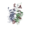 4c7lSC S: Starting model for refinement C: citing same article ( |
|---|---|
| Similar structure data |


-Links
 PDBj
PDBj


- Assembly
| Deposited unit | 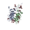
| ||||||||
|---|---|---|---|---|---|---|---|---|---|
| 1 |
| ||||||||
| Unit cell |
|
-Components
-Protein , 1 types, 2 molecules AB
| #1: Protein | Mass: 43503.355 Da / Num. of mol.: 2 / Fragment: RESIDUES 25-403 / Mutation: YES Source method: isolated from a genetically manipulated source Source: (gene. exp.) MURINE HEPATITIS VIRUS / Strain: S / Plasmid: PCD5- MHV-S-HE-T-FC / Cell line (production host): HEK293S / Production host:  HOMO SAPIENS (human) HOMO SAPIENS (human)References: UniProt: O55252, UniProt: P31614*PLUS, sialate O-acetylesterase |
|---|
-Sugars , 5 types, 14 molecules 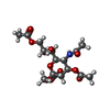

| #2: Polysaccharide | 2-acetamido-2-deoxy-beta-D-glucopyranose-(1-4)-2-acetamido-2-deoxy-beta-D-glucopyranose Source method: isolated from a genetically manipulated source #3: Polysaccharide | Source method: isolated from a genetically manipulated source #4: Polysaccharide | alpha-D-mannopyranose-(1-3)-[alpha-D-mannopyranose-(1-6)]alpha-D-mannopyranose-(1-6)-[alpha-D- ...alpha-D-mannopyranose-(1-3)-[alpha-D-mannopyranose-(1-6)]alpha-D-mannopyranose-(1-6)-[alpha-D-mannopyranose-(1-3)]beta-D-mannopyranose-(1-4)-2-acetamido-2-deoxy-beta-D-glucopyranose-(1-4)-2-acetamido-2-deoxy-beta-D-glucopyranose | Source method: isolated from a genetically manipulated source #5: Sugar |  Type: D-saccharide, alpha linking / Mass: 407.370 Da / Num. of mol.: 2 Type: D-saccharide, alpha linking / Mass: 407.370 Da / Num. of mol.: 2Source method: isolated from a genetically manipulated source Formula: C16H25NO11 #7: Sugar | ChemComp-NAG /  Type: D-saccharide, beta linking / Mass: 221.208 Da / Num. of mol.: 4 Type: D-saccharide, beta linking / Mass: 221.208 Da / Num. of mol.: 4Source method: isolated from a genetically manipulated source Formula: C8H15NO6 |
|---|
-Non-polymers , 3 types, 78 molecules 


| #6: Chemical |  Mass: 39.098 Da / Num. of mol.: 2 / Source method: obtained synthetically / Formula: K Mass: 39.098 Da / Num. of mol.: 2 / Source method: obtained synthetically / Formula: K#8: Chemical | ChemComp-GOL / |  Mass: 92.094 Da / Num. of mol.: 1 / Source method: obtained synthetically / Formula: C3H8O3 Mass: 92.094 Da / Num. of mol.: 1 / Source method: obtained synthetically / Formula: C3H8O3#9: Water | ChemComp-HOH / | Mass: 18.015 Da / Num. of mol.: 75 / Source method: isolated from a natural source / Formula: H2O |
|---|
-Details
| Has protein modification | Y |
|---|
-Experimental details
-Experiment
| Experiment | Method: X-RAY DIFFRACTION / Number of used crystals: 1 |
|---|
- Sample preparation
| Crystal | Density Matthews: 3.2 Å3/Da / Density % sol: 61 % / Description: NONE |
|---|---|
| Crystal grow | pH: 3.6 Details: 0.2 M KH2PO4, 0.2 M SODIUM MALONATE, 15% (W/V) PEG3350 AND 5% (W/V) GLYCEROL, pH 3.6 |
-Data collection
| Diffraction | Mean temperature: 100 K |
|---|---|
| Diffraction source | Source: SYNCHROTRON / Site: ESRF  / Beamline: ID23-2 / Wavelength: 0.8726 / Beamline: ID23-2 / Wavelength: 0.8726 |
| Detector | Type: MARRESEARCH / Detector: CCD / Date: Mar 11, 2007 / Details: MIRRORS |
| Radiation | Protocol: SINGLE WAVELENGTH / Monochromatic (M) / Laue (L): M / Scattering type: x-ray |
| Radiation wavelength | Wavelength: 0.8726 Å / Relative weight: 1 |
| Reflection | Resolution: 2.5→54.5 Å / Num. obs: 46670 / % possible obs: 100 % / Observed criterion σ(I): 2 / Redundancy: 7.4 % / Rmerge(I) obs: 0.12 / Net I/σ(I): 12.7 |
| Reflection shell | Resolution: 2.5→2.64 Å / Redundancy: 7.4 % / Rmerge(I) obs: 0.9 / Mean I/σ(I) obs: 2.1 / % possible all: 100 |
- Processing
| Software |
| ||||||||||||||||||||||||||||||||||||||||||||||||||||||||||||||||||||||||||||||||||||||||||||||||||||||||||||||||||||||||||||||||||||||||||||||||||||||||||||||||||||||||||||||||||||||
|---|---|---|---|---|---|---|---|---|---|---|---|---|---|---|---|---|---|---|---|---|---|---|---|---|---|---|---|---|---|---|---|---|---|---|---|---|---|---|---|---|---|---|---|---|---|---|---|---|---|---|---|---|---|---|---|---|---|---|---|---|---|---|---|---|---|---|---|---|---|---|---|---|---|---|---|---|---|---|---|---|---|---|---|---|---|---|---|---|---|---|---|---|---|---|---|---|---|---|---|---|---|---|---|---|---|---|---|---|---|---|---|---|---|---|---|---|---|---|---|---|---|---|---|---|---|---|---|---|---|---|---|---|---|---|---|---|---|---|---|---|---|---|---|---|---|---|---|---|---|---|---|---|---|---|---|---|---|---|---|---|---|---|---|---|---|---|---|---|---|---|---|---|---|---|---|---|---|---|---|---|---|---|---|
| Refinement | Method to determine structure: MOLECULAR REPLACEMENT Starting model: PDB ENTRY 4C7L Resolution: 2.5→83.92 Å / Cor.coef. Fo:Fc: 0.929 / Cor.coef. Fo:Fc free: 0.907 / SU B: 14.042 / SU ML: 0.161 / Cross valid method: THROUGHOUT / ESU R: 0.294 / ESU R Free: 0.232 / Stereochemistry target values: MAXIMUM LIKELIHOOD Details: HYDROGENS HAVE BEEN ADDED IN THE RIDING POSITIONS. U VALUES WITH TLS ADDED
| ||||||||||||||||||||||||||||||||||||||||||||||||||||||||||||||||||||||||||||||||||||||||||||||||||||||||||||||||||||||||||||||||||||||||||||||||||||||||||||||||||||||||||||||||||||||
| Solvent computation | Ion probe radii: 0.8 Å / Shrinkage radii: 0.8 Å / VDW probe radii: 1.4 Å / Solvent model: BABINET MODEL WITH MASK | ||||||||||||||||||||||||||||||||||||||||||||||||||||||||||||||||||||||||||||||||||||||||||||||||||||||||||||||||||||||||||||||||||||||||||||||||||||||||||||||||||||||||||||||||||||||
| Displacement parameters | Biso mean: 45.024 Å2
| ||||||||||||||||||||||||||||||||||||||||||||||||||||||||||||||||||||||||||||||||||||||||||||||||||||||||||||||||||||||||||||||||||||||||||||||||||||||||||||||||||||||||||||||||||||||
| Refinement step | Cycle: LAST / Resolution: 2.5→83.92 Å
| ||||||||||||||||||||||||||||||||||||||||||||||||||||||||||||||||||||||||||||||||||||||||||||||||||||||||||||||||||||||||||||||||||||||||||||||||||||||||||||||||||||||||||||||||||||||
| Refine LS restraints |
|