 Movie
Movie Controller

[English] 日本語
 Yorodumi
- PDB-4c1s: Glycoside hydrolase family 76 (mannosidase) Bt3792 from Bacteroid... -
+ Open data

- Basic information
| Entry | Database: PDB / ID: 4c1s | ||||||
|---|---|---|---|---|---|---|---|
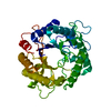
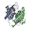
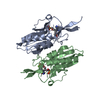
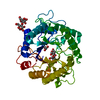
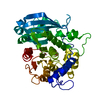
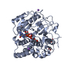
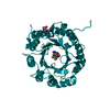
| Title | Glycoside hydrolase family 76 (mannosidase) Bt3792 from Bacteroides thetaiotaomicron VPI-5482 | ||||||
 Components Components | GLYCOSIDE HYDROLASE FAMILY 76 MANNOSIDASE | ||||||
 Keywords Keywords | HYDROLASE / GUT MICROBIOTA | ||||||
| Function / homology |  Function and homology information Function and homology information | ||||||
| Biological species |  BACTEROIDES THETAIOTAOMICRON VPI-5482 (bacteria) BACTEROIDES THETAIOTAOMICRON VPI-5482 (bacteria) | ||||||
| Method |  X-RAY DIFFRACTION / SYNCHROTRON / MOLECULAR REPLACEMENT / Resolution: 2.1 Å X-RAY DIFFRACTION / SYNCHROTRON / MOLECULAR REPLACEMENT / Resolution: 2.1 Å | ||||||
 Authors Authors | Cuskin, F. / Lowe, E.C. / Zhu, Y. / Temple, M. / Thompson, A.J. / Cartmell, A. / Piens, K. / Bracke, D. / Vervecken, W. / Munoz-Munoz, J.L. ...Cuskin, F. / Lowe, E.C. / Zhu, Y. / Temple, M. / Thompson, A.J. / Cartmell, A. / Piens, K. / Bracke, D. / Vervecken, W. / Munoz-Munoz, J.L. / Suits, M.D.L. / Boraston, A.B. / Williams, S.J. / Davies, G.J. / Abbott, W.D. / Martens, E.C. / Gilbert, H.J. | ||||||
 Citation Citation | Journal: Nature / Year: 2015 Title: Human Gut Bacteroidetes Can Utilize Yeast Mannan Through a Selfish Mechanism. Authors: Cuskin, F. / Lowe, E.C. / Temple, M.J. / Zhu, Y. / Cameron, E.A. / Pudlo, N.A. / Porter, N.T. / Urs, K. / Thompson, A.J. / Cartmell, A. / Rogowski, A. / Hamilton, B.S. / Chen, R. / Tolbert, ...Authors: Cuskin, F. / Lowe, E.C. / Temple, M.J. / Zhu, Y. / Cameron, E.A. / Pudlo, N.A. / Porter, N.T. / Urs, K. / Thompson, A.J. / Cartmell, A. / Rogowski, A. / Hamilton, B.S. / Chen, R. / Tolbert, T.J. / Piens, K. / Bracke, D. / Vervecken, W. / Hakki, Z. / Speciale, G. / Munoz-Munoz, J.L. / Day, A. / Pena, M.J. / Mclean, R. / Suits, M.D. / Boraston, A.B. / Atherly, T. / Ziemer, C.J. / Williams, S.J. / Davies, G.J. / Abbott, D.W. / Martens, E.C. / Gilbert, H.J. | ||||||
| History |
|
- Structure visualization
| Structure viewer | Molecule: MolmilJmol/JSmol |
|---|
- Downloads & links
-Download
| PDBx/mmCIF format | 4c1s.cif.gz | 169.2 KB | Display | PDBx/mmCIF format |
|---|---|---|---|---|
| PDB format | pdb4c1s.ent.gz | 133.5 KB | Display | PDB format |
| PDBx/mmJSON format | 4c1s.json.gz | Tree view | PDBx/mmJSON format | |
| Others |  Other downloads Other downloads |
-Validation report
| Arichive directory | https://data.pdbj.org/pub/pdb/validation_reports/c1/4c1sftp://data.pdbj.org/pub/pdb/validation_reports/c1/4c1s | HTTPS FTP |
|---|
-Related structure data
| Related structure data |  4c1rC 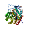 4utfC 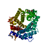 3k7xS C: citing same article ( S: Starting model for refinement |
|---|---|
| Similar structure data |


-Links
 PDBj
PDBj
- Assembly
| Deposited unit | 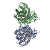
| ||||||||
|---|---|---|---|---|---|---|---|---|---|
| 1 | 
| ||||||||
| 2 | 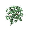
| ||||||||
| Unit cell |
|
-Components
| #1: Protein | Mass: 43430.590 Da / Num. of mol.: 2 / Fragment: ENZYMATIC FRAGMENT, RESIDUES 155-525 Source method: isolated from a genetically manipulated source Details: BT3792 GENE FRAGMENT ENCODING RESIDUES 155- 514 Source: (gene. exp.) BACTEROIDES THETAIOTAOMICRON VPI-5482 (bacteria)Gene: BT3792 / Production host: References: UniProt: Q8A174, mannan endo-1,6-alpha-mannosidase #2: Chemical | ChemComp-EDO /   Mass: 62.068 Da / Num. of mol.: 13 / Source method: obtained synthetically / Formula: C2H6O2 Mass: 62.068 Da / Num. of mol.: 13 / Source method: obtained synthetically / Formula: C2H6O2#3: Chemical |   Mass: 92.094 Da / Num. of mol.: 2 / Source method: obtained synthetically / Formula: C3H8O3 Mass: 92.094 Da / Num. of mol.: 2 / Source method: obtained synthetically / Formula: C3H8O3#4: Water | ChemComp-HOH / |  Mass: 18.015 Da / Num. of mol.: 287 / Source method: isolated from a natural source / Formula: H2O Mass: 18.015 Da / Num. of mol.: 287 / Source method: isolated from a natural source / Formula: H2O |
|---|
-Experimental details
-Experiment
| Experiment | Method: X-RAY DIFFRACTION / Number of used crystals: 1 |
|---|
- Sample preparation
| Crystal | Density Matthews: 2.7 Å3/Da / Density % sol: 54 % / Description: NONE |
|---|---|
| Crystal grow | pH: 8 Details: BT3792 AT A 20 MG PER ML BUFFERED IN IN 25 MM TRIS-HCL (PH 8.0), 500 MM NACL, 2 MM DTT WITH A RESERVOIR SOLUTION CONSISTING OF 5% GLYCEROL (V/V), 24% (W PER V) POLYETHYLENE GLYCOL 2,000 ...Details: BT3792 AT A 20 MG PER ML BUFFERED IN IN 25 MM TRIS-HCL (PH 8.0), 500 MM NACL, 2 MM DTT WITH A RESERVOIR SOLUTION CONSISTING OF 5% GLYCEROL (V/V), 24% (W PER V) POLYETHYLENE GLYCOL 2,000 MONOMETHYL ETHER, 0.25 M SODIUM ACETATE, AND BIS-TRIS-HCL (PH 5.5) |
-Data collection
| Diffraction | Mean temperature: 100 K |
|---|---|
| Diffraction source | Source: SYNCHROTRON / Site: CLSI  / Beamline: 08ID-1 / Wavelength: 0.98005 / Beamline: 08ID-1 / Wavelength: 0.98005 |
| Detector | Type: MARRESEARCH MX-300HE / Detector: CCD / Date: Sep 14, 2012 Details: COLLIMATING MIRROR WITH TWO STRIPES (SI, RH AND PT) , TOROIDAL FOCUSING MIRROR (RH AND PT) |
| Radiation | Monochromator: KOHZU DOUBLE CRYSTAL MONOCHROMATOR (DCM), FEATURING INDIRECTLY WATER- COOLED FIRST CRYSTAL AND FLAT, LONG SECOND CRYSTAL Protocol: SINGLE WAVELENGTH / Monochromatic (M) / Laue (L): M / Scattering type: x-ray |
| Radiation wavelength | Wavelength: 0.98005 Å / Relative weight: 1 |
| Reflection | Resolution: 2.1→48 Å / Num. obs: 52497 / % possible obs: 99.9 % / Observed criterion σ(I): 0 / Redundancy: 4.2 % / Rmerge(I) obs: 0.11 / Net I/σ(I): 10.4 |
| Reflection shell | Resolution: 2.1→2.21 Å / Redundancy: 4.2 % / Rmerge(I) obs: 0.48 / Mean I/σ(I) obs: 3.3 / % possible all: 99.9 |
- Processing
| Software |
| ||||||||||||||||||||||||||||||||||||||||||||||||||||||||||||||||||||||||||||||||||||||||||||||||||||||||||||||||||||||||||||||||||||||||||||||||||||||||||||||||||||||||||||||||||||||
|---|---|---|---|---|---|---|---|---|---|---|---|---|---|---|---|---|---|---|---|---|---|---|---|---|---|---|---|---|---|---|---|---|---|---|---|---|---|---|---|---|---|---|---|---|---|---|---|---|---|---|---|---|---|---|---|---|---|---|---|---|---|---|---|---|---|---|---|---|---|---|---|---|---|---|---|---|---|---|---|---|---|---|---|---|---|---|---|---|---|---|---|---|---|---|---|---|---|---|---|---|---|---|---|---|---|---|---|---|---|---|---|---|---|---|---|---|---|---|---|---|---|---|---|---|---|---|---|---|---|---|---|---|---|---|---|---|---|---|---|---|---|---|---|---|---|---|---|---|---|---|---|---|---|---|---|---|---|---|---|---|---|---|---|---|---|---|---|---|---|---|---|---|---|---|---|---|---|---|---|---|---|---|---|
| Refinement | Method to determine structure: MOLECULAR REPLACEMENT Starting model: PDB ENTRY 3K7X Resolution: 2.1→48.03 Å / Cor.coef. Fo:Fc: 0.932 / Cor.coef. Fo:Fc free: 0.904 / SU B: 3.911 / SU ML: 0.108 / Cross valid method: THROUGHOUT / ESU R: 0.05 / ESU R Free: 0.041 / Stereochemistry target values: MAXIMUM LIKELIHOOD Details: HYDROGENS HAVE BEEN ADDED IN THE RIDING POSITIONS. U VALUES REFINED INDIVIDUALLY
| ||||||||||||||||||||||||||||||||||||||||||||||||||||||||||||||||||||||||||||||||||||||||||||||||||||||||||||||||||||||||||||||||||||||||||||||||||||||||||||||||||||||||||||||||||||||
| Solvent computation | Ion probe radii: 0.8 Å / Shrinkage radii: 0.8 Å / VDW probe radii: 1.2 Å / Solvent model: MASK | ||||||||||||||||||||||||||||||||||||||||||||||||||||||||||||||||||||||||||||||||||||||||||||||||||||||||||||||||||||||||||||||||||||||||||||||||||||||||||||||||||||||||||||||||||||||
| Displacement parameters | Biso mean: 24.111 Å2
| ||||||||||||||||||||||||||||||||||||||||||||||||||||||||||||||||||||||||||||||||||||||||||||||||||||||||||||||||||||||||||||||||||||||||||||||||||||||||||||||||||||||||||||||||||||||
| Refinement step | Cycle: LAST / Resolution: 2.1→48.03 Å
| ||||||||||||||||||||||||||||||||||||||||||||||||||||||||||||||||||||||||||||||||||||||||||||||||||||||||||||||||||||||||||||||||||||||||||||||||||||||||||||||||||||||||||||||||||||||
| Refine LS restraints |
|