 Movie
Movie Controller

[English] 日本語
 Yorodumi
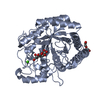
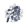
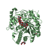
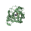
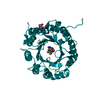
- PDB-1r8l: The structure of endo-beta-1,4-galactanase from Bacillus licheniformis -
+ Open data

- Basic information
| Entry | Database: PDB / ID: 1r8l | ||||||
|---|---|---|---|---|---|---|---|
| Title | The structure of endo-beta-1,4-galactanase from Bacillus licheniformis | ||||||
 Components Components | endo-beta-1,4-galactanase | ||||||
 Keywords Keywords | HYDROLASE / (beta-alpha)8-barrel / calcium ion / glycosyl hydrolase / family 53 / clan GH-A | ||||||
| Function / homology |  Function and homology information Function and homology informationarabinogalactan endo-beta-1,4-galactanase / arabinogalactan endo-1,4-beta-galactosidase activity / glucosidase activity / pectin catabolic process / Hydrolases; Glycosylases; Glycosidases, i.e. enzymes that hydrolyse O- and S-glycosyl compounds / metal ion binding Similarity search - Function | ||||||
| Biological species |  | ||||||
| Method |  X-RAY DIFFRACTION / MOLECULAR REPLACEMENT / Resolution: 2.6 Å X-RAY DIFFRACTION / MOLECULAR REPLACEMENT / Resolution: 2.6 Å | ||||||
 Authors Authors | Ryttersgaard, C. / Le Nours, J. / Lo Leggio, L. / Jorgensen, C.T. / Christensen, L.L. / Bjornvad, M. / Larsen, S. | ||||||
 Citation Citation | Journal: J.Mol.Biol. / Year: 2004 Title: The structure of endo-beta-1,4-galactanase from Bacillus licheniformis in complex with two oligosaccharide products Authors: Ryttersgaard, C. / Le Nours, J. / Lo Leggio, L. / Jorgensen, C.T. / Christensen, L.L. / Bjornvad, M. / Larsen, S. | ||||||
| History |
|
- Structure visualization
| Structure viewer | Molecule: MolmilJmol/JSmol |
|---|
- Downloads & links
-Download
| PDBx/mmCIF format | 1r8l.cif.gz | 159 KB | Display | PDBx/mmCIF format |
|---|---|---|---|---|
| PDB format | pdb1r8l.ent.gz | 125.6 KB | Display | PDB format |
| PDBx/mmJSON format | 1r8l.json.gz | Tree view | PDBx/mmJSON format | |
| Others |  Other downloads Other downloads |
-Validation report
| Arichive directory | https://data.pdbj.org/pub/pdb/validation_reports/r8/1r8lftp://data.pdbj.org/pub/pdb/validation_reports/r8/1r8l | HTTPS FTP |
|---|
-Related structure data
| Related structure data | 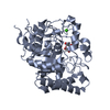 1ur0C  1ur4C  1fhlS S: Starting model for refinement C: citing same article ( |
|---|---|
| Similar structure data |


-Links
 PDBj
PDBj- Assembly
| Deposited unit | 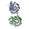
| ||||||||
|---|---|---|---|---|---|---|---|---|---|
| 1 | 
| ||||||||
| 2 | 
| ||||||||
| Unit cell |
| ||||||||
| Details | The biological assembly is a monomer of either chain A or B |
-Components
| #1: Protein | Mass: 43778.434 Da / Num. of mol.: 2 Source method: isolated from a genetically manipulated source Source: (gene. exp.) References: UniProt: Q65CX5, arabinogalactan endo-beta-1,4-galactanase #2: Chemical |   Mass: 40.078 Da / Num. of mol.: 2 / Source method: obtained synthetically / Formula: Ca Mass: 40.078 Da / Num. of mol.: 2 / Source method: obtained synthetically / Formula: Ca#3: Water | ChemComp-HOH / |  Mass: 18.015 Da / Num. of mol.: 124 / Source method: isolated from a natural source / Formula: H2O Mass: 18.015 Da / Num. of mol.: 124 / Source method: isolated from a natural source / Formula: H2O |
|---|
-Experimental details
-Experiment
| Experiment | Method: X-RAY DIFFRACTION / Number of used crystals: 1 |
|---|
- Sample preparation
| Crystal | Density Matthews: 2.45 Å3/Da / Density % sol: 49.76 % |
|---|---|
| Crystal grow | Temperature: 293 K / Method: vapor diffusion, hanging drop / pH: 5 Details: PEG 1500, sodium acetate, pH 5.0, VAPOR DIFFUSION, HANGING DROP, temperature 293K |
-Data collection
| Diffraction | Mean temperature: 293 K |
|---|---|
| Diffraction source | Source: ROTATING ANODE / Type: RIGAKU RU200 / Wavelength: 1.5418 Å |
| Detector | Type: RIGAKU RAXIS IIC / Detector: IMAGE PLATE / Date: Sep 1, 2000 |
| Radiation | Protocol: SINGLE WAVELENGTH / Monochromatic (M) / Laue (L): M / Scattering type: x-ray |
| Radiation wavelength | Wavelength: 1.5418 Å / Relative weight: 1 |
| Reflection | Resolution: 2.6→19.67 Å / Num. all: 25224 / Num. obs: 25224 / % possible obs: 96.5 % / Observed criterion σ(F): 0 / Observed criterion σ(I): 0 / Redundancy: 2.3 % / Biso Wilson estimate: 38.871 Å2 / Rmerge(I) obs: 0.101 / Net I/σ(I): 8.61 |
| Reflection shell | Resolution: 2.6→2.64 Å / Redundancy: 2.3 % / Rmerge(I) obs: 0.41 / Mean I/σ(I) obs: 2 / Num. unique all: 1239 / % possible all: 95.5 |
- Processing
| Software |
| |||||||||||||||||||||||||
|---|---|---|---|---|---|---|---|---|---|---|---|---|---|---|---|---|---|---|---|---|---|---|---|---|---|---|
| Refinement | Method to determine structure: MOLECULAR REPLACEMENT Starting model: PDN ENTRY 1FHL Resolution: 2.6→19.67 Å / Isotropic thermal model: RESTRAINED / Cross valid method: THROUGHOUT / σ(F): 0 / Stereochemistry target values: Engh & Huber
| |||||||||||||||||||||||||
| Displacement parameters | Biso mean: 31.7 Å2 | |||||||||||||||||||||||||
| Refine analyze |
| |||||||||||||||||||||||||
| Refinement step | Cycle: LAST / Resolution: 2.6→19.67 Å
| |||||||||||||||||||||||||
| Refine LS restraints |
| |||||||||||||||||||||||||
| LS refinement shell | Resolution: 2.6→2.76 Å / Rfactor Rfree error: 0.016
|
