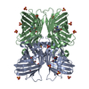
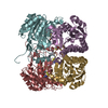
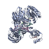
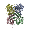
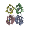
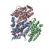
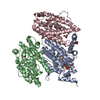
[English] 日本語
Yorodumi
- PDB-4c0s: Mammalian translation elongation factor eEF1A2 -

+
Open data


ID or keywords:

Loading...

-
Basic information

Entry
Database: PDB / ID: 4c0s
TitleMammalian translation elongation factor eEF1A2
ComponentsELONGATION FACTOR 1-ALPHA 2
KeywordsTRANSLATION
Function / homology
Function and homology information


translation elongation factor activity / GTPase activity / GTP binding / nucleus
Similarity search - Function
Translation elongation factor EF1A, eukaryotic/archaeal / Translation elongation factor EFTu/EF1A, C-terminal / Elongation factor Tu C-terminal domain / Translation elongation factor EF1A/initiation factor IF2gamma, C-terminal / Translation factors / Tr-type G domain, conserved site / Translational (tr)-type guanine nucleotide-binding (G) domain signature. / Translation elongation factor EFTu-like, domain 2 / Elongation factor Tu domain 2 / Translational (tr)-type GTP-binding domain ...Translation elongation factor EF1A, eukaryotic/archaeal / Translation elongation factor EFTu/EF1A, C-terminal / Elongation factor Tu C-terminal domain / Translation elongation factor EF1A/initiation factor IF2gamma, C-terminal / Translation factors / Tr-type G domain, conserved site / Translational (tr)-type guanine nucleotide-binding (G) domain signature. / Translation elongation factor EFTu-like, domain 2 / Elongation factor Tu domain 2 / Translational (tr)-type GTP-binding domain / Elongation factor Tu GTP binding domain / Translational (tr)-type guanine nucleotide-binding (G) domain profile. / Elongation Factor Tu (Ef-tu); domain 3 / Translation protein, beta-barrel domain superfamily / P-loop containing nucleotide triphosphate hydrolases / Beta Barrel / P-loop containing nucleoside triphosphate hydrolase / Rossmann fold / 3-Layer(aba) Sandwich / Mainly Beta / Alpha Beta
Similarity search - Domain/homology
GUANOSINE-5'-DIPHOSPHATE / Elongation factor 1-alpha 2
Similarity search - Component
Biological speciesORYCTOLAGUS CUNICULUS (rabbit)
MethodX-RAY DIFFRACTION / SYNCHROTRON / MIR / Resolution: 2.703 Å
AuthorsCrepin, T. / Shalak, V.F. / Yaremchuk, A.D. / Vlasenko, D.O. / McCarthy, A.A. / Negrutskii, B.S. / Tukalo, M.A. / El'skaya, A.V.
CitationJournal: Nucleic Acids Res. / Year: 2014
Title: Mammalian Translation Elongation Factor Eef1A2: X-Ray Structure and New Features of Gdp/GTP Exchange Mechanism in Higher Eukaryotes
Authors: Crepin, T. / Mccarthy, A. / Negrutskii, A. / Shalak, V.F. / Tukalo, V. / Vlasenko, D.O. / Yaremchuk, A.D.
History
DepositionAug 7, 2013Deposition site: PDBE / Processing site: PDBE
Revision 1.0Aug 27, 2014Provider: repository / Type: Initial release
Revision 1.1Oct 29, 2014Group: Database references
Revision 1.2Nov 26, 2014Group: Database references
Revision 1.3Jan 25, 2017Group: Data collection
Remark 700 SHEET DETERMINATION METHOD: DSSP THE SHEETS PRESENTED AS "AC" IN EACH CHAIN ON SHEET RECORDS BELOW ... SHEET DETERMINATION METHOD: DSSP THE SHEETS PRESENTED AS "AC" IN EACH CHAIN ON SHEET RECORDS BELOW IS ACTUALLY AN 6-STRANDED BARREL THIS IS REPRESENTED BY A 7-STRANDED SHEET IN WHICH THE FIRST AND LAST STRANDS ARE IDENTICAL. THE SHEETS PRESENTED AS "AD" IN EACH CHAIN ON SHEET RECORDS BELOW IS ACTUALLY AN 6-STRANDED BARREL THIS IS REPRESENTED BY A 7-STRANDED SHEET IN WHICH THE FIRST AND LAST STRANDS ARE IDENTICAL. THE SHEETS PRESENTED AS "BC" IN EACH CHAIN ON SHEET RECORDS BELOW IS ACTUALLY AN 6-STRANDED BARREL THIS IS REPRESENTED BY A 7-STRANDED SHEET IN WHICH THE FIRST AND LAST STRANDS ARE IDENTICAL. THE SHEETS PRESENTED AS "BD" IN EACH CHAIN ON SHEET RECORDS BELOW IS ACTUALLY AN -9-STRANDED BARREL THIS IS REPRESENTED BY A -8-STRANDED SHEET IN WHICH THE FIRST AND LAST STRANDS ARE IDENTICAL.

-
Structure visualization

Structure viewerMolecule:
MolmilJmol/JSmol

Downloads & links

-
Assembly

Deposited unit
A: ELONGATION FACTOR 1-ALPHA 2
B: ELONGATION FACTOR 1-ALPHA 2
hetero molecules


Theoretical massNumber of molelcules
Total (without water)102,3456
Polymers101,4102
Non-polymers9354
Water1629
1


  • Idetical with deposited unit
  • defined by author&software
TypeNameSymmetry operationNumber
identity operation1_555x,y,z1
Buried area6310 Å2
ΔGint-28.5 kcal/mol
Surface area35990 Å2
MethodPISA
Unit cell
Length a, b, c (Å)135.370, 135.370, 304.620
Angle α, β, γ (deg.)90.00, 90.00, 120.00
Int Tables number178
Space group name H-MP6122

-
Components

#1: Protein ELONGATION FACTOR 1-ALPHA 2 / EF-1-ALPHA-2 / EUKARYOTIC ELONGATION FACTOR 1 A-2 / EEF1A-2 / STATIN-S1


Mass: 50705.059 Da / Num. of mol.: 2 / Source method: isolated from a natural source / Source: (natural) ORYCTOLAGUS CUNICULUS (rabbit) / Tissue: MUSCLESkeletal muscle / References: UniProt: Q71V39
#2: Chemical ChemComp-GDP / GUANOSINE-5'-DIPHOSPHATE / Guanosine diphosphate


Type: RNA linking / Mass: 443.201 Da / Num. of mol.: 2 / Source method: obtained synthetically / Formula: C10H15N5O11P2 / Comment: GDP, energy-carrying molecule*YM
#3: Chemical ChemComp-MG / MAGNESIUM ION


Mass: 24.305 Da / Num. of mol.: 2 / Source method: obtained synthetically / Formula: Mg
#4: Water ChemComp-HOH / water / Water


Mass: 18.015 Da / Num. of mol.: 9 / Source method: isolated from a natural source / Formula: H2O

-
Experimental details

-
Experiment

ExperimentMethod: X-RAY DIFFRACTION / Number of used crystals: 10

-
Sample preparation

CrystalDensity Matthews: 3.87 Å3/Da / Density % sol: 68.1 % / Description: NONE
Crystal growDetails: 50 MM MES PH 5.6, 2.4 M AMMONIUM SULFATE, 10 MM MAGNESIUM ACETATE

-
Data collection

DiffractionMean temperature: 100 K
Diffraction sourceSource: SYNCHROTRON / Site: ESRF / Beamline: ID14-4 / Wavelength: 0.97936
DetectorType: ADSC CCD / Detector: CCD / Date: Dec 18, 2010
RadiationProtocol: SINGLE WAVELENGTH / Monochromatic (M) / Laue (L): M / Scattering type: x-ray
Radiation wavelengthWavelength: 0.97936 Å / Relative weight: 1
ReflectionResolution: 2.7→25 Å / Num. obs: 45528 / % possible obs: 98.4 % / Observed criterion σ(I): 2.5 / Redundancy: 7.5 % / Rmerge(I) obs: 0.08 / Net I/σ(I): 18.52
Reflection shellResolution: 2.7→2.8 Å / Redundancy: 7.7 % / Rmerge(I) obs: 0.86 / Mean I/σ(I) obs: 2.76 / % possible all: 95.6

-
Processing

Software
NameVersionClassification
REFMAC5.6.0114refinement
XDSdata reduction
XSCALEdata scaling
RefinementMethod to determine structure: MIR
Starting model: NONE

Resolution: 2.703→25 Å / Cor.coef. Fo:Fc: 0.947 / Cor.coef. Fo:Fc free: 0.916 / SU B: 11.093 / SU ML: 0.22 / Cross valid method: THROUGHOUT / σ(F): 2.703 / ESU R: 0.374 / ESU R Free: 0.283 / Stereochemistry target values: MAXIMUM LIKELIHOOD / Details: HYDROGENS HAVE BEEN ADDED IN THE RIDING POSITIONS.
RfactorNum. reflection% reflection
Rfree0.2556 2302 5.16 %
Rwork0.2019 --
obs0.205 45527 98.972 %
Solvent computationIon probe radii: 0.8 Å / VDW probe radii: 1.2 Å / Solvent model: MASK BULK SOLVENT
Displacement parametersBiso mean: 62.295 Å2
Baniso -1Baniso -2Baniso -3
1-1.623 Å20.811 Å20 Å2
2--1.623 Å20 Å2
3----2.434 Å2
Refinement stepCycle: LAST / Resolution: 2.703→25 Å
ProteinNucleic acidLigandSolventTotal
Num. atoms6884 0 58 9 6951
Refine LS restraints
Refine-IDTypeDev idealDev ideal targetNumber
X-RAY DIFFRACTIONr_bond_refined_d0.0150.0197085
X-RAY DIFFRACTIONr_bond_other_d
X-RAY DIFFRACTIONr_angle_refined_deg1.5121.9839601
X-RAY DIFFRACTIONr_angle_other_deg
X-RAY DIFFRACTIONr_dihedral_angle_1_deg8.965890
X-RAY DIFFRACTIONr_dihedral_angle_2_deg39.33524.42276
X-RAY DIFFRACTIONr_dihedral_angle_3_deg22.713151255
X-RAY DIFFRACTIONr_dihedral_angle_4_deg19.9191536
X-RAY DIFFRACTIONr_chiral_restr0.1170.21075
X-RAY DIFFRACTIONr_gen_planes_refined0.010.0215210
X-RAY DIFFRACTIONr_gen_planes_other
X-RAY DIFFRACTIONr_nbd_refined0.2630.23366
X-RAY DIFFRACTIONr_nbd_other
X-RAY DIFFRACTIONr_nbtor_refined0.3310.24774
X-RAY DIFFRACTIONr_nbtor_other
X-RAY DIFFRACTIONr_xyhbond_nbd_refined0.1720.2263
X-RAY DIFFRACTIONr_xyhbond_nbd_other
X-RAY DIFFRACTIONr_metal_ion_refined
X-RAY DIFFRACTIONr_metal_ion_other
X-RAY DIFFRACTIONr_symmetry_vdw_refined
X-RAY DIFFRACTIONr_symmetry_vdw_other
X-RAY DIFFRACTIONr_symmetry_hbond_refined
X-RAY DIFFRACTIONr_symmetry_hbond_other
X-RAY DIFFRACTIONr_symmetry_metal_ion_refined
X-RAY DIFFRACTIONr_symmetry_metal_ion_other
X-RAY DIFFRACTIONr_mcbond_it6.3066.1827084
X-RAY DIFFRACTIONr_mcbond_other
X-RAY DIFFRACTIONr_mcangle_it9.3339.1569600
X-RAY DIFFRACTIONr_mcangle_other
X-RAY DIFFRACTIONr_scbond_it14.17218.8932457
X-RAY DIFFRACTIONr_scbond_other
X-RAY DIFFRACTIONr_scangle_it12.24129.4568281
X-RAY DIFFRACTIONr_scangle_other
X-RAY DIFFRACTIONr_long_range_B_refined
X-RAY DIFFRACTIONr_long_range_B_other
X-RAY DIFFRACTIONr_rigid_bond_restr
X-RAY DIFFRACTIONr_sphericity_free
X-RAY DIFFRACTIONr_sphericity_bonded
LS refinement shellResolution: 2.703→2.772 Å / Total num. of bins used: 20
RfactorNum. reflection% reflection
Rfree0.422 144 -
Rwork0.344 2806 -
obs--98.761 %

+
About Yorodumi

-
News

-
Feb 9, 2022. New format data for meta-information of EMDB entries

New format data for meta-information of EMDB entries

  • Version 3 of the EMDB header file is now the official format.
  • The previous official version 1.9 will be removed from the archive.

Related info.:EMDB header

External links:wwPDB to switch to version 3 of the EMDB data model

-
Aug 12, 2020. Covid-19 info

Covid-19 info

URL: https://pdbj.org/emnavi/covid19.php

New page: Covid-19 featured information page in EM Navigator.

Related info.:Covid-19 info / Mar 5, 2020. Novel coronavirus structure data

+
Mar 5, 2020. Novel coronavirus structure data

Novel coronavirus structure data

Related info.:Yorodumi Speices / Aug 12, 2020. Covid-19 info

External links:COVID-19 featured content - PDBj / Molecule of the Month (242):Coronavirus Proteases

+
Jan 31, 2019. EMDB accession codes are about to change! (news from PDBe EMDB page)

EMDB accession codes are about to change! (news from PDBe EMDB page)

  • The allocation of 4 digits for EMDB accession codes will soon come to an end. Whilst these codes will remain in use, new EMDB accession codes will include an additional digit and will expand incrementally as the available range of codes is exhausted. The current 4-digit format prefixed with “EMD-” (i.e. EMD-XXXX) will advance to a 5-digit format (i.e. EMD-XXXXX), and so on. It is currently estimated that the 4-digit codes will be depleted around Spring 2019, at which point the 5-digit format will come into force.
  • The EM Navigator/Yorodumi systems omit the EMD- prefix.

Related info.:Q: What is EMD? / ID/Accession-code notation in Yorodumi/EM Navigator

External links:EMDB Accession Codes are Changing Soon! / Contact to PDBj

+
Jul 12, 2017. Major update of PDB

Major update of PDB

  • wwPDB released updated PDB data conforming to the new PDBx/mmCIF dictionary.
  • This is a major update changing the version number from 4 to 5, and with Remediation, in which all the entries are updated.
  • In this update, many items about electron microscopy experimental information are reorganized (e.g. em_software).
  • Now, EM Navigator and Yorodumi are based on the updated data.

External links:wwPDB Remediation / Enriched Model Files Conforming to OneDep Data Standards Now Available in the PDB FTP Archive

-
Yorodumi

Thousand views of thousand structures

  • Yorodumi is a browser for structure data from EMDB, PDB, SASBDB, etc.
  • This page is also the successor to EM Navigator detail page, and also detail information page/front-end page for Omokage search.
  • The word "yorodu" (or yorozu) is an old Japanese word meaning "ten thousand". "mi" (miru) is to see.

Related info.:EMDB / PDB / SASBDB / Comparison of 3 databanks / Yorodumi Search / Aug 31, 2016. New EM Navigator & Yorodumi / Yorodumi Papers / Jmol/JSmol / Function and homology information / Changes in new EM Navigator and Yorodumi

Read more