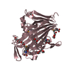
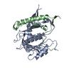
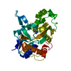
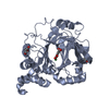
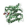
[English] 日本語
Yorodumi
- PDB-4bzg: Crystal structure of galactose mutarotase GalM from Bacillus subt... -

+
Open data


ID or keywords:

Loading...

-
Basic information

Entry
Database: PDB / ID: 4bzg
TitleCrystal structure of galactose mutarotase GalM from Bacillus subtilis in complex with maltose
ComponentsALDOSE 1-EPIMERASE
KeywordsISOMERASE
Function / homology
Function and homology information


aldose 1-epimerase / aldose 1-epimerase activity / galactose catabolic process via UDP-galactose / glucose metabolic process / carbohydrate binding / cytoplasm
Similarity search - Function
Aldose 1-/Glucose-6-phosphate 1-epimerase / Aldose 1-epimerase / Beta-galactosidase; Chain A, domain 5 - #10 / Glycoside hydrolase-type carbohydrate-binding / Beta-galactosidase; Chain A, domain 5 / Galactose mutarotase-like domain superfamily / Distorted Sandwich / Mainly Beta
Similarity search - Domain/homology
alpha-maltose / CITRIC ACID / Aldose 1-epimerase
Similarity search - Component
Biological speciesBACILLUS SUBTILIS SUBSP. SUBTILIS STR. 168 (bacteria)
MethodX-RAY DIFFRACTION / MOLECULAR REPLACEMENT / Resolution: 2.13 Å
AuthorsVanden Broeck, A. / Sauvage, E. / Herman, R. / Kerff, F. / Duez, C. / Charlier, P.
CitationJournal: To be Published
Title: Crystal Structure of Galactose Mutarotase Galm from Bacillus Subtilis in Complex with Maltose
Authors: Vanden Broeck, A. / Sauvage, E. / Herman, R. / Kerff, F. / Duez, C. / Charlier, P.
History
DepositionJul 25, 2013Deposition site: PDBE / Processing site: PDBE
Revision 1.0Aug 13, 2014Provider: repository / Type: Initial release
Revision 2.0Jul 29, 2020Group: Atomic model / Data collection ...Atomic model / Data collection / Derived calculations / Non-polymer description / Other / Structure summary
Category: atom_site / chem_comp ...atom_site / chem_comp / entity / entity_name_com / pdbx_branch_scheme / pdbx_chem_comp_identifier / pdbx_database_status / pdbx_entity_branch / pdbx_entity_branch_descriptor / pdbx_entity_branch_link / pdbx_entity_branch_list / pdbx_entity_nonpoly / pdbx_molecule_features / pdbx_nonpoly_scheme / struct_conn / struct_site / struct_site_gen
Item: _atom_site.B_iso_or_equiv / _atom_site.Cartn_x ..._atom_site.B_iso_or_equiv / _atom_site.Cartn_x / _atom_site.Cartn_y / _atom_site.Cartn_z / _atom_site.auth_asym_id / _atom_site.auth_atom_id / _atom_site.auth_comp_id / _atom_site.auth_seq_id / _atom_site.label_atom_id / _atom_site.label_comp_id / _chem_comp.formula / _chem_comp.formula_weight / _chem_comp.id / _chem_comp.mon_nstd_flag / _chem_comp.name / _chem_comp.type / _entity.formula_weight / _entity.pdbx_description / _entity.type / _pdbx_database_status.status_code_sf
Description: Carbohydrate remediation / Provider: repository / Type: Remediation
Revision 2.1Dec 20, 2023Group: Data collection / Database references ...Data collection / Database references / Refinement description / Structure summary
Category: chem_comp / chem_comp_atom ...chem_comp / chem_comp_atom / chem_comp_bond / database_2 / pdbx_initial_refinement_model
Item: _chem_comp.pdbx_synonyms / _database_2.pdbx_DOI / _database_2.pdbx_database_accession

-
Structure visualization

Structure viewerMolecule:
MolmilJmol/JSmol

Downloads & links

-
Assembly

Deposited unit
A: ALDOSE 1-EPIMERASE
B: ALDOSE 1-EPIMERASE
hetero molecules


Theoretical massNumber of molelcules
Total (without water)75,1645
Polymers74,2872
Non-polymers8773
Water5,657314
1
A: ALDOSE 1-EPIMERASE
hetero molecules


Theoretical massNumber of molelcules
Total (without water)37,4862
Polymers37,1441
Non-polymers3421
Water181
TypeNameSymmetry operationNumber
identity operation1_555x,y,z1
MethodPISA
2
B: ALDOSE 1-EPIMERASE
hetero molecules


Theoretical massNumber of molelcules
Total (without water)37,6783
Polymers37,1441
Non-polymers5342
Water181
TypeNameSymmetry operationNumber
identity operation1_555x,y,z1
MethodPISA
Unit cell
Length a, b, c (Å)82.461, 84.202, 125.445
Angle α, β, γ (deg.)90.00, 90.00, 90.00
Int Tables number18
Space group name H-MP21212

-
Components

#1: Protein ALDOSE 1-EPIMERASE / GALACTOSE MUTAROTASE


Mass: 37143.719 Da / Num. of mol.: 2
Source method: isolated from a genetically manipulated source
Source: (gene. exp.) BACILLUS SUBTILIS SUBSP. SUBTILIS STR. 168 (bacteria)
Gene: GALM / Production host: ESCHERICHIA COLI K-12 (bacteria) / Strain (production host): KRX / References: UniProt: P39840, aldose 1-epimerase
#2: Polysaccharide alpha-D-glucopyranose-(1-4)-alpha-D-glucopyranose / alpha-maltose


Type: oligosaccharide, Oligosaccharide / Class: Nutrient / Mass: 342.297 Da / Num. of mol.: 2
Source method: isolated from a genetically manipulated source
Details: oligosaccharide / References: alpha-maltose
DescriptorTypeProgram
DGlcpa1-4DGlcpa1-ROHGlycam Condensed SequenceGMML 1.0
WURCS=2.0/1,2,1/[a2122h-1a_1-5]/1-1/a4-b1WURCSPDB2Glycan 1.1.0
[][a-D-Glcp]{[(4+1)][a-D-Glcp]{}}LINUCSPDB-CARE
#3: Chemical ChemComp-CIT / CITRIC ACID


Mass: 192.124 Da / Num. of mol.: 1 / Source method: obtained synthetically / Formula: C6H8O7
#4: Water ChemComp-HOH / water


Mass: 18.015 Da / Num. of mol.: 314 / Source method: isolated from a natural source / Formula: H2O

-
Experimental details

-
Experiment

ExperimentMethod: X-RAY DIFFRACTION / Number of used crystals: 1

-
Sample preparation

CrystalDensity Matthews: 2.78 Å3/Da / Density % sol: 55.41 % / Description: NONE

-
Data collection

DiffractionMean temperature: 100 K
Diffraction sourceSource: ROTATING ANODE / Wavelength: 1.5418
DetectorType: MAR scanner 345 mm plate / Detector: IMAGE PLATE / Date: Dec 3, 2012
RadiationProtocol: SINGLE WAVELENGTH / Monochromatic (M) / Laue (L): M / Scattering type: x-ray
Radiation wavelengthWavelength: 1.5418 Å / Relative weight: 1
ReflectionResolution: 2.13→29.67 Å / Num. obs: 49195 / % possible obs: 98.5 % / Observed criterion σ(I): 0 / Redundancy: 7 % / Rmerge(I) obs: 0.11 / Net I/σ(I): 13.2
Reflection shellResolution: 2.13→2.24 Å / Redundancy: 5.9 % / Rmerge(I) obs: 0.57 / Mean I/σ(I) obs: 3.4 / % possible all: 89.8

-
Processing

Software
NameVersionClassification
REFMAC5.6.0117refinement
XDSdata reduction
XDSdata scaling
PHASERphasing
RefinementMethod to determine structure: MOLECULAR REPLACEMENT
Starting model: PDB ENTRY 3MWX
Resolution: 2.13→29.67 Å / Cor.coef. Fo:Fc: 0.955 / Cor.coef. Fo:Fc free: 0.934 / SU B: 9.015 / SU ML: 0.121 / Cross valid method: THROUGHOUT / ESU R: 0.191 / ESU R Free: 0.171 / Stereochemistry target values: MAXIMUM LIKELIHOOD / Details: HYDROGENS HAVE BEEN ADDED IN THE RIDING POSITIONS.
RfactorNum. reflection% reflectionSelection details
Rfree0.22671 2457 5 %RANDOM
Rwork0.18284 ---
obs0.18504 46673 98.41 %-
Solvent computationIon probe radii: 0.8 Å / Shrinkage radii: 0.8 Å / VDW probe radii: 1.2 Å / Solvent model: MASK
Displacement parametersBiso mean: 31.681 Å2
Baniso -1Baniso -2Baniso -3
1--0.06 Å20 Å20 Å2
2--0.11 Å20 Å2
3----0.06 Å2
Refinement stepCycle: LAST / Resolution: 2.13→29.67 Å
ProteinNucleic acidLigandSolventTotal
Num. atoms5222 0 59 314 5595
Refine LS restraints
Refine-IDTypeDev idealDev ideal targetNumber
X-RAY DIFFRACTIONr_bond_refined_d0.0130.025431
X-RAY DIFFRACTIONr_bond_other_d
X-RAY DIFFRACTIONr_angle_refined_deg1.5671.9487398
X-RAY DIFFRACTIONr_angle_other_deg
X-RAY DIFFRACTIONr_dihedral_angle_1_deg7.0075643
X-RAY DIFFRACTIONr_dihedral_angle_2_deg40.92824.791263
X-RAY DIFFRACTIONr_dihedral_angle_3_deg14.53915891
X-RAY DIFFRACTIONr_dihedral_angle_4_deg25.2431516
X-RAY DIFFRACTIONr_chiral_restr0.1070.2822
X-RAY DIFFRACTIONr_gen_planes_refined0.0070.0214128
X-RAY DIFFRACTIONr_gen_planes_other
X-RAY DIFFRACTIONr_nbd_refined
X-RAY DIFFRACTIONr_nbd_other
X-RAY DIFFRACTIONr_nbtor_refined
X-RAY DIFFRACTIONr_nbtor_other
X-RAY DIFFRACTIONr_xyhbond_nbd_refined
X-RAY DIFFRACTIONr_xyhbond_nbd_other
X-RAY DIFFRACTIONr_metal_ion_refined
X-RAY DIFFRACTIONr_metal_ion_other
X-RAY DIFFRACTIONr_symmetry_vdw_refined
X-RAY DIFFRACTIONr_symmetry_vdw_other
X-RAY DIFFRACTIONr_symmetry_hbond_refined
X-RAY DIFFRACTIONr_symmetry_hbond_other
X-RAY DIFFRACTIONr_symmetry_metal_ion_refined
X-RAY DIFFRACTIONr_symmetry_metal_ion_other
X-RAY DIFFRACTIONr_mcbond_it
X-RAY DIFFRACTIONr_mcbond_other
X-RAY DIFFRACTIONr_mcangle_it
X-RAY DIFFRACTIONr_mcangle_other
X-RAY DIFFRACTIONr_scbond_it
X-RAY DIFFRACTIONr_scbond_other
X-RAY DIFFRACTIONr_scangle_it
X-RAY DIFFRACTIONr_scangle_other
X-RAY DIFFRACTIONr_long_range_B_refined
X-RAY DIFFRACTIONr_long_range_B_other
X-RAY DIFFRACTIONr_rigid_bond_restr
X-RAY DIFFRACTIONr_sphericity_free
X-RAY DIFFRACTIONr_sphericity_bonded
LS refinement shellResolution: 2.125→2.18 Å / Total num. of bins used: 20
RfactorNum. reflection% reflection
Rfree0.389 136 -
Rwork0.3 2579 -
obs--78.49 %
Refinement TLS params.

Method: refined / Refine-ID: X-RAY DIFFRACTION

IDL112)L122)L132)L222)L232)L332)S11 (Å °)S12 (Å °)S13 (Å °)S21 (Å °)S22 (Å °)S23 (Å °)S31 (Å °)S32 (Å °)S33 (Å °)T112)T122)T132)T222)T232)T332)Origin x (Å)Origin y (Å)Origin z (Å)
10.53650.189-0.09330.6533-0.13680.49190.0390.0336-0.101-0.0427-0.0232-0.10310.00890.0841-0.01580.07680.02240.01490.0368-0.00560.1362-1.76316.229-13.749
20.65330.3475-0.31450.5621-0.16290.43480.01580.1183-0.1253-0.07280.0024-0.05210.0365-0.005-0.01820.10330.0176-0.00210.0495-0.01730.1194-12.11411.063-16.593
30.7099-0.01340.04940.53620.12670.47-0.0059-0.03090.03190.03620.00730.03730.03350.0005-0.00140.08330.00680.00750.0420.01020.0971-13.35821.489-2.266
42.8548-1.091-0.05371.737-0.32930.69110.1631-0.22640.09420.1083-0.11170.0548-0.04330.0164-0.05140.0891-0.00410.01470.0733-0.01020.0683-11.36127.7694.467
50.66540.0722-0.13610.34780.12430.31160.0102-0.0112-0.016-0.0062-0.0097-0.0118-0.0164-0.0097-0.00050.08150.00640.00320.04980.01470.1264-12.90522.58-5.16
627.00513.640616.338815.20280.208217.671-0.13731.67910.74350.40910.26310.9937-0.56311.6037-0.12590.1435-0.04880.14130.1742-0.04160.23260.54937.014-9.444
70.07010.3115-0.02671.88950.48082.2958-0.02480.04620.00940.0310.01020.1687-0.0113-0.2850.01460.0836-0.02890.05280.1564-0.01450.0715-3.26122.983-45.317
83.17660.065-1.44360.52170.01260.6762-0.23950.1075-0.1779-0.14940.1198-0.05220.1584-0.04340.11970.2922-0.00970.13170.084-0.05170.105712.8219.594-53.559
90.4463-0.0416-0.81861.68040.41621.5753-0.27710.2023-0.1497-0.26480.0628-0.34670.4402-0.37340.21430.3063-0.07080.12930.1601-0.11010.138116.6244.007-55.848
100.8932-1.0549-1.52622.90631.71514.5154-0.0925-0.1744-0.0890.05820.1397-0.0148-0.08980.1872-0.04720.1061-0.01950.03630.09310.02910.05656.32222.602-36.984
110.8718-0.1955-0.15670.9773-0.23360.3413-0.06630.11350.0255-0.13680.0001-0.14970.0661-0.02170.06620.1282-0.00590.06090.1303-0.00730.042314.68621.636-57.117
1238.26596.450630.11331.08865.078623.7021-3.027-0.83953.7227-0.5078-0.02650.6365-2.4275-0.64353.05350.72180.1094-0.12950.1623-0.04020.4331-1.90440.159-57.286
Refinement TLS group
IDRefine-IDRefine TLS-IDAuth asym-IDAuth seq-ID
1X-RAY DIFFRACTION1A2 - 85
2X-RAY DIFFRACTION2A86 - 153
3X-RAY DIFFRACTION3A154 - 219
4X-RAY DIFFRACTION4A220 - 243
5X-RAY DIFFRACTION5A244 - 318
6X-RAY DIFFRACTION6A319 - 323
7X-RAY DIFFRACTION7B2 - 52
8X-RAY DIFFRACTION8B53 - 85
9X-RAY DIFFRACTION9B86 - 105
10X-RAY DIFFRACTION10B106 - 124
11X-RAY DIFFRACTION11B125 - 319
12X-RAY DIFFRACTION12B320 - 324

+
About Yorodumi

-
News

-
Feb 9, 2022. New format data for meta-information of EMDB entries

New format data for meta-information of EMDB entries

  • Version 3 of the EMDB header file is now the official format.
  • The previous official version 1.9 will be removed from the archive.

Related info.:EMDB header

External links:wwPDB to switch to version 3 of the EMDB data model

-
Aug 12, 2020. Covid-19 info

Covid-19 info

URL: https://pdbjlvh1.pdbj.org/emnavi/covid19.php

New page: Covid-19 featured information page in EM Navigator.

Related info.:Covid-19 info / Mar 5, 2020. Novel coronavirus structure data

+
Mar 5, 2020. Novel coronavirus structure data

Novel coronavirus structure data

Related info.:Yorodumi Speices / Aug 12, 2020. Covid-19 info

External links:COVID-19 featured content - PDBj / Molecule of the Month (242):Coronavirus Proteases

+
Jan 31, 2019. EMDB accession codes are about to change! (news from PDBe EMDB page)

EMDB accession codes are about to change! (news from PDBe EMDB page)

  • The allocation of 4 digits for EMDB accession codes will soon come to an end. Whilst these codes will remain in use, new EMDB accession codes will include an additional digit and will expand incrementally as the available range of codes is exhausted. The current 4-digit format prefixed with “EMD-” (i.e. EMD-XXXX) will advance to a 5-digit format (i.e. EMD-XXXXX), and so on. It is currently estimated that the 4-digit codes will be depleted around Spring 2019, at which point the 5-digit format will come into force.
  • The EM Navigator/Yorodumi systems omit the EMD- prefix.

Related info.:Q: What is EMD? / ID/Accession-code notation in Yorodumi/EM Navigator

External links:EMDB Accession Codes are Changing Soon! / Contact to PDBj

+
Jul 12, 2017. Major update of PDB

Major update of PDB

  • wwPDB released updated PDB data conforming to the new PDBx/mmCIF dictionary.
  • This is a major update changing the version number from 4 to 5, and with Remediation, in which all the entries are updated.
  • In this update, many items about electron microscopy experimental information are reorganized (e.g. em_software).
  • Now, EM Navigator and Yorodumi are based on the updated data.

External links:wwPDB Remediation / Enriched Model Files Conforming to OneDep Data Standards Now Available in the PDB FTP Archive

-
Yorodumi

Thousand views of thousand structures

  • Yorodumi is a browser for structure data from EMDB, PDB, SASBDB, etc.
  • This page is also the successor to EM Navigator detail page, and also detail information page/front-end page for Omokage search.
  • The word "yorodu" (or yorozu) is an old Japanese word meaning "ten thousand". "mi" (miru) is to see.

Related info.:EMDB / PDB / SASBDB / Comparison of 3 databanks / Yorodumi Search / Aug 31, 2016. New EM Navigator & Yorodumi / Yorodumi Papers / Jmol/JSmol / Function and homology information / Changes in new EM Navigator and Yorodumi

Read more