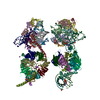
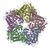
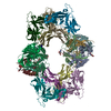
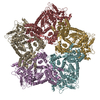
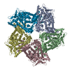
Journal: Structure / Year: 2013 Title: Plate tectonics of virus shell assembly and reorganization in phage φ8, a distant relative of mammalian reoviruses. Authors: Kamel El Omari / Geoff Sutton / Janne J Ravantti / Hanwen Zhang / Thomas S Walter / Jonathan M Grimes / Dennis H Bamford / David I Stuart / Erika J Mancini / Abstract: The hallmark of a virus is its capsid, which harbors the viral genome and is formed from protein subunits, which assemble following precise geometric rules. dsRNA viruses use an unusual protein ...The hallmark of a virus is its capsid, which harbors the viral genome and is formed from protein subunits, which assemble following precise geometric rules. dsRNA viruses use an unusual protein multiplicity (120 copies) to form their closed capsids. We have determined the atomic structure of the capsid protein (P1) from the dsRNA cystovirus Φ8. In the crystal P1 forms pentamers, very similar in shape to facets of empty procapsids, suggesting an unexpected assembly pathway that proceeds via a pentameric intermediate. Unlike the elongated proteins used by dsRNA mammalian reoviruses, P1 has a compact trapezoid-like shape and a distinct arrangement in the shell, with two near-identical conformers in nonequivalent structural environments. Nevertheless, structural similarity with the analogous protein from the mammalian viruses suggests a common ancestor. The unusual shape of the molecule may facilitate dramatic capsid expansion during phage maturation, allowing P1 to switch interaction interfaces to provide capsid plasticity.
Protocol: SINGLE WAVELENGTH / Monochromatic (M) / Laue (L): M / Scattering type: x-ray
Radiation wavelength
Wavelength: 0.9326 Å / Relative weight: 1
Reflection
Resolution: 3.7→30 Å / Num. obs: 271956 / % possible obs: 97.1 % / Observed criterion σ(I): 2.1 / Redundancy: 11.1 % / Rmerge(I) obs: 0.17 / Net I/σ(I): 15.1
Reflection shell
Resolution: 3.7→3.83 Å / Redundancy: 4.8 % / Rmerge(I) obs: 1 / Mean I/σ(I) obs: 2.1 / % possible all: 71.3
-
Processing
Software
Name
Version
Classification
REFMAC
5.7.0032
refinement
HKL-2000
datareduction
SCALEPACK
datascaling
HKL2Map
phasing
Refinement
Method to determine structure: SAD Starting model: NONE Resolution: 3.7→30.01 Å / Cor.coef. Fo:Fc: 0.925 / Cor.coef. Fo:Fc free: 0.919 / SU B: 28.249 / SU ML: 0.387 / Cross valid method: THROUGHOUT / ESU R: 1.372 / ESU R Free: 0.465 / Stereochemistry target values: MAXIMUM LIKELIHOOD Details: HYDROGENS HAVE BEEN ADDED IN THE RIDING POSITIONS. U VALUES REFINED INDIVIDUALLY
Rfactor
Num. reflection
% reflection
Selection details
Rfree
0.25836
13628
5 %
RANDOM
Rwork
0.24448
-
-
-
obs
0.24519
256979
96.96 %
-
Solvent computation
Ion probe radii: 0.8 Å / Shrinkage radii: 0.8 Å / VDW probe radii: 1.2 Å / Solvent model: MASK
 Movie
Movie Controller


 Open data
 Basic information
 Components
 Keywords
 Function and homology information
 Pseudomonas phage phi8 (virus)
 X-RAY DIFFRACTION /
  Authors
 Citation
 Structure visualization
 Downloads & links
 Other downloads
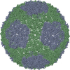


 PDBj
PDBj Assembly

 Sample preparation
 / Beamline: ID14-2 / Wavelength: 0.9326
  Processing