 Movie
Movie Controller


+ Open data

- Basic information
| Entry | Database: PDB / ID: 4bmg | ||||||
|---|---|---|---|---|---|---|---|
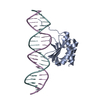
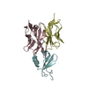
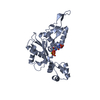
| Title | Crystal structure of hexameric HBc149 Y132A | ||||||
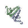
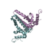
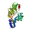
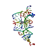
 Components Components | CAPSID PROTEIN | ||||||
 Keywords Keywords | VIRAL PROTEIN / PROTEIN FOLDING / ALLOSTERY | ||||||
| Function / homology |  Function and homology information Function and homology informationmicrotubule-dependent intracellular transport of viral material towards nucleus / T=4 icosahedral viral capsid / viral penetration into host nucleus / host cell / host cell cytoplasm / symbiont entry into host cell / structural molecule activity / DNA binding / RNA binding / identical protein binding Similarity search - Function | ||||||
| Biological species |   HEPATITIS B VIRUS HEPATITIS B VIRUS | ||||||
| Method |  X-RAY DIFFRACTION / SYNCHROTRON / MOLECULAR REPLACEMENT / Resolution: 3 Å X-RAY DIFFRACTION / SYNCHROTRON / MOLECULAR REPLACEMENT / Resolution: 3 Å | ||||||
 Authors Authors | Juergens, M.C. / Alexander, C.G. / Shepherd, D.A. / Ashcroft, A.E. / Ferguson, N. | ||||||
 Citation Citation | Journal: Proc.Natl.Acad.Sci.USA / Year: 2013 Title: Thermodynamic Origins of Protein Folding, Allostery and Capsid Formation in the Human Hepatitis B Virus Core Protein Authors: Alexander, C.G. / Juergens, M.C. / Shepherd, D.A. / Freund, S. / Ashcroft, A.E. / Ferguson, N. | ||||||
| History |
|
- Structure visualization
| Structure viewer | Molecule: MolmilJmol/JSmol |
|---|
- Downloads & links
-Download
| PDBx/mmCIF format | 4bmg.cif.gz | 341.4 KB | Display | PDBx/mmCIF format |
|---|---|---|---|---|
| PDB format | pdb4bmg.ent.gz | 286.2 KB | Display | PDB format |
| PDBx/mmJSON format | 4bmg.json.gz | Tree view | PDBx/mmJSON format | |
| Others |  Other downloads Other downloads |
-Validation report
| Summary document | 4bmg_validation.pdf.gz | 477 KB | Display | wwPDB validaton report |
|---|---|---|---|---|
| Full document | 4bmg_full_validation.pdf.gz | 493.3 KB | Display | |
| Data in XML | 4bmg_validation.xml.gz | 31.2 KB | Display | |
| Data in CIF | 4bmg_validation.cif.gz | 42.4 KB | Display | |
| Arichive directory | https://data.pdbj.org/pub/pdb/validation_reports/bm/4bmgftp://data.pdbj.org/pub/pdb/validation_reports/bm/4bmg | HTTPS FTP |
-Related structure data
| Related structure data |  3kxsS S: Starting model for refinement |
|---|---|
| Similar structure data |

-Links
 PDBj
PDBj

- Assembly
| Deposited unit | 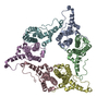
| ||||||||||||||||||||||||
|---|---|---|---|---|---|---|---|---|---|---|---|---|---|---|---|---|---|---|---|---|---|---|---|---|---|
| 1 | 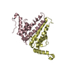
| ||||||||||||||||||||||||
| 2 | 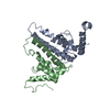
| ||||||||||||||||||||||||
| 3 | 
| ||||||||||||||||||||||||
| Unit cell |
| ||||||||||||||||||||||||
| Noncrystallographic symmetry (NCS) | NCS oper:
|
-Components
| #1: Protein | Mass: 17358.785 Da / Num. of mol.: 6 / Fragment: RESIDUES 1-149 / Mutation: YES Source method: isolated from a genetically manipulated source Details: DISULFIDE LINK BETWEEN C61 RESIDUES AT THE DIMER INTERFACE Source: (gene. exp.) HEPATITIS B VIRUS / Strain: ADYW / Production host:  Has protein modification | Y | |
|---|
-Experimental details
-Experiment
| Experiment | Method: X-RAY DIFFRACTION / Number of used crystals: 1 |
|---|
- Sample preparation
| Crystal | Density Matthews: 3.22 Å3/Da / Density % sol: 61.8 % / Description: NONE |
|---|---|
| Crystal grow | Temperature: 293 K / pH: 5.5 Details: 100 MM CITRATE PH 5.0, 15 % (V/V) ISO-PROPANOL, 1% (W/V) PEG 10 000, 293 K |
-Data collection
| Diffraction | Mean temperature: 93 K |
|---|---|
| Diffraction source | Source: SYNCHROTRON / Site: SLS  / Beamline: X06DA / Wavelength: 0.98 / Beamline: X06DA / Wavelength: 0.98 |
| Detector | Type: DECTRIS PILATUS 2M-F / Detector: PIXEL / Date: Feb 6, 2013 / Details: TOROIDAL MIRROR (M2) |
| Radiation | Monochromator: DCCM / Protocol: SINGLE WAVELENGTH / Monochromatic (M) / Laue (L): M / Scattering type: x-ray |
| Radiation wavelength | Wavelength: 0.98 Å / Relative weight: 1 |
| Reflection | Resolution: 3→48.69 Å / Num. obs: 24745 / % possible obs: 95.3 % / Observed criterion σ(I): 1.8 / Redundancy: 1.8 % / Biso Wilson estimate: 79.77 Å2 / Rmerge(I) obs: 0.03 / Net I/σ(I): 17.2 |
| Reflection shell | Resolution: 3→3.08 Å / Redundancy: 1.7 % / Rmerge(I) obs: 0.43 / Mean I/σ(I) obs: 1.8 / % possible all: 94 |
- Processing
| Software |
| ||||||||||||||||||||||||||||||||||||||||||||||||||||||||||||||||||||||
|---|---|---|---|---|---|---|---|---|---|---|---|---|---|---|---|---|---|---|---|---|---|---|---|---|---|---|---|---|---|---|---|---|---|---|---|---|---|---|---|---|---|---|---|---|---|---|---|---|---|---|---|---|---|---|---|---|---|---|---|---|---|---|---|---|---|---|---|---|---|---|---|
| Refinement | Method to determine structure: MOLECULAR REPLACEMENT Starting model: PDB ENTRY 3KXS Resolution: 3→44.526 Å / SU ML: 0.5 / σ(F): 1.97 / Phase error: 37.63 / Stereochemistry target values: ML
| ||||||||||||||||||||||||||||||||||||||||||||||||||||||||||||||||||||||
| Solvent computation | Shrinkage radii: 0.9 Å / VDW probe radii: 1.11 Å / Solvent model: FLAT BULK SOLVENT MODEL | ||||||||||||||||||||||||||||||||||||||||||||||||||||||||||||||||||||||
| Displacement parameters | Biso mean: 96.54 Å2 | ||||||||||||||||||||||||||||||||||||||||||||||||||||||||||||||||||||||
| Refinement step | Cycle: LAST / Resolution: 3→44.526 Å
| ||||||||||||||||||||||||||||||||||||||||||||||||||||||||||||||||||||||
| Refine LS restraints |
| ||||||||||||||||||||||||||||||||||||||||||||||||||||||||||||||||||||||
| LS refinement shell |
| ||||||||||||||||||||||||||||||||||||||||||||||||||||||||||||||||||||||
| Refinement TLS params. | Method: refined / Origin x: 37.4836 Å / Origin y: 2.0907 Å / Origin z: 19.9676 Å
| ||||||||||||||||||||||||||||||||||||||||||||||||||||||||||||||||||||||
| Refinement TLS group | Selection details: ALL |
