 Movie
Movie Controller

[English] 日本語
 Yorodumi
- PDB-3kxs: Crystal structure of HBV capsid mutant dimer (oxy form), strain adyw -
+ Open data

- Basic information
| Entry | Database: PDB / ID: 3kxs | ||||||
|---|---|---|---|---|---|---|---|
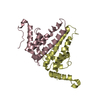
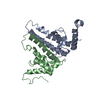
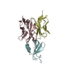
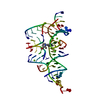
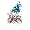
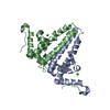
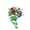
| Title | Crystal structure of HBV capsid mutant dimer (oxy form), strain adyw | ||||||
 Components Components | Capsid protein | ||||||
 Keywords Keywords | VIRAL PROTEIN / Core protein / capsid / hepadnavirus / four helix bundle / icosahedral / dimer / Capsid protein / DNA-binding / Host cytoplasm / Host-virus interaction / Phosphoprotein / RNA-binding / Virion | ||||||
| Function / homology |  Function and homology information Function and homology informationmicrotubule-dependent intracellular transport of viral material towards nucleus / T=4 icosahedral viral capsid / viral penetration into host nucleus / host cell / host cell cytoplasm / symbiont entry into host cell / structural molecule activity / DNA binding / RNA binding / identical protein binding Similarity search - Function | ||||||
| Biological species |  Hepatitis B virus subtype adyw Hepatitis B virus subtype adyw | ||||||
| Method |  X-RAY DIFFRACTION / MOLECULAR REPLACEMENT / Resolution: 2.25 Å X-RAY DIFFRACTION / MOLECULAR REPLACEMENT / Resolution: 2.25 Å | ||||||
 Authors Authors | Packianathan, C. / Katen, S.P. / Zlotnick, A. | ||||||
 Citation Citation | Journal: J.Virol. / Year: 2010 Title: Conformational changes in the hepatitis B virus core protein are consistent with a role for allostery in virus assembly Authors: Packianathan, C. / Katen, S.P. / Dann, C.E. / Zlotnick, A. #1: Journal: Biochemistry / Year: 2009 Title: A Mutant Hepatitis B Virus Core Protein Mimics Inhibitors of Icosahedral Capsid Self-Assembly Authors: Bourne, C.R. / Katen, S.P. / Fulz, M.R. / Packianathan, C. / Zlotnick, A. | ||||||
| History |
|
- Structure visualization
| Structure viewer | Molecule: MolmilJmol/JSmol |
|---|
- Downloads & links
-Download
| PDBx/mmCIF format | 3kxs.cif.gz | 176 KB | Display | PDBx/mmCIF format |
|---|---|---|---|---|
| PDB format | pdb3kxs.ent.gz | 142.7 KB | Display | PDB format |
| PDBx/mmJSON format | 3kxs.json.gz | Tree view | PDBx/mmJSON format | |
| Others |  Other downloads Other downloads |
-Validation report
| Arichive directory | https://data.pdbj.org/pub/pdb/validation_reports/kx/3kxsftp://data.pdbj.org/pub/pdb/validation_reports/kx/3kxs | HTTPS FTP |
|---|
-Related structure data
| Related structure data | 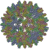 1qgtS S: Starting model for refinement |
|---|---|
| Similar structure data |


-Links
 PDBj
PDBj

- Assembly
| Deposited unit | 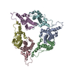
| ||||||||
|---|---|---|---|---|---|---|---|---|---|
| 1 | 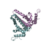
| ||||||||
| 2 | 
| ||||||||
| 3 | 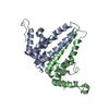
| ||||||||
| Unit cell |
| ||||||||
| Details | Authors state that the mutant is an assembly deficient dimer of core protein. The wt is also a dimer in solution but can be assembled into an icosahedron (PDB accession 2G33). |
-Components
| #1: Protein | Mass: 16065.363 Da / Num. of mol.: 6 / Fragment: assembly domain residues 1 to 149 / Mutation: Y132A Source method: isolated from a genetically manipulated source Source: (gene. exp.) Hepatitis B virus subtype adyw / Genus: orthohepadnavirus / Species: Hepatitis B virus / Strain: ADYW / Gene: C / Plasmid: pET11c / Production host:  #2: Water | ChemComp-HOH / |  Mass: 18.015 Da / Num. of mol.: 208 / Source method: isolated from a natural source / Formula: H2O Mass: 18.015 Da / Num. of mol.: 208 / Source method: isolated from a natural source / Formula: H2OHas protein modification | Y | |
|---|
-Experimental details
-Experiment
| Experiment | Method: X-RAY DIFFRACTION / Number of used crystals: 1 |
|---|
- Sample preparation
| Crystal | Density Matthews: 2.82 Å3/Da / Density % sol: 56.45 % |
|---|---|
| Crystal grow | Temperature: 293 K / Method: vapor diffusion, hanging drop / pH: 6.5 Details: PEG1500, Calcium acetate, sodium Cacodylate, pH 6.5, VAPOR DIFFUSION, HANGING DROP, temperature 293K |
-Data collection
| Diffraction | Mean temperature: 93 K |
|---|---|
| Diffraction source | Source: ROTATING ANODE / Type: RIGAKU RUH3R / Wavelength: 1.5418 Å |
| Detector | Type: RIGAKU RAXIS IV++ / Detector: IMAGE PLATE / Date: Jul 18, 2009 / Details: mirror |
| Radiation | Monochromator: Osmic Blue confocal mirror / Protocol: SINGLE WAVELENGTH / Monochromatic (M) / Laue (L): M / Scattering type: x-ray |
| Radiation wavelength | Wavelength: 1.5418 Å / Relative weight: 1 |
| Reflection | Resolution: 2.25→50 Å / Num. all: 228368 / Num. obs: 228368 / % possible obs: 99.9 % / Observed criterion σ(F): 0 / Observed criterion σ(I): 0 / Redundancy: 4.5 % / Rmerge(I) obs: 0.076 / Net I/σ(I): 41.8 |
| Reflection shell | Resolution: 2.25→2.33 Å / Redundancy: 4.4 % / Rmerge(I) obs: 0.572 / Mean I/σ(I) obs: 5.34 / % possible all: 100 |
- Processing
| Software |
| ||||||||||||||||||||||||||||||||||||||||||||||||||||||||||||||||||||||||||||||||||||||||||||||||||||||||||||||||||||||||||||||||||||||||||||||||||||||||||||||||||||||||||
|---|---|---|---|---|---|---|---|---|---|---|---|---|---|---|---|---|---|---|---|---|---|---|---|---|---|---|---|---|---|---|---|---|---|---|---|---|---|---|---|---|---|---|---|---|---|---|---|---|---|---|---|---|---|---|---|---|---|---|---|---|---|---|---|---|---|---|---|---|---|---|---|---|---|---|---|---|---|---|---|---|---|---|---|---|---|---|---|---|---|---|---|---|---|---|---|---|---|---|---|---|---|---|---|---|---|---|---|---|---|---|---|---|---|---|---|---|---|---|---|---|---|---|---|---|---|---|---|---|---|---|---|---|---|---|---|---|---|---|---|---|---|---|---|---|---|---|---|---|---|---|---|---|---|---|---|---|---|---|---|---|---|---|---|---|---|---|---|---|---|---|---|
| Refinement | Method to determine structure: MOLECULAR REPLACEMENT Starting model: PDB entry 1QGT Resolution: 2.25→25.32 Å / Cor.coef. Fo:Fc: 0.949 / Cor.coef. Fo:Fc free: 0.931 / Isotropic thermal model: isotropic / Cross valid method: THROUGHOUT / ESU R: 0.294 / ESU R Free: 0.231 / Stereochemistry target values: MAXIMUM LIKELIHOOD
| ||||||||||||||||||||||||||||||||||||||||||||||||||||||||||||||||||||||||||||||||||||||||||||||||||||||||||||||||||||||||||||||||||||||||||||||||||||||||||||||||||||||||||
| Solvent computation | Ion probe radii: 0.8 Å / Shrinkage radii: 0.8 Å / VDW probe radii: 1.4 Å / Solvent model: MASK | ||||||||||||||||||||||||||||||||||||||||||||||||||||||||||||||||||||||||||||||||||||||||||||||||||||||||||||||||||||||||||||||||||||||||||||||||||||||||||||||||||||||||||
| Displacement parameters | Biso mean: 48.36 Å2
| ||||||||||||||||||||||||||||||||||||||||||||||||||||||||||||||||||||||||||||||||||||||||||||||||||||||||||||||||||||||||||||||||||||||||||||||||||||||||||||||||||||||||||
| Refinement step | Cycle: LAST / Resolution: 2.25→25.32 Å
| ||||||||||||||||||||||||||||||||||||||||||||||||||||||||||||||||||||||||||||||||||||||||||||||||||||||||||||||||||||||||||||||||||||||||||||||||||||||||||||||||||||||||||
| Refine LS restraints |
| ||||||||||||||||||||||||||||||||||||||||||||||||||||||||||||||||||||||||||||||||||||||||||||||||||||||||||||||||||||||||||||||||||||||||||||||||||||||||||||||||||||||||||
| LS refinement shell | Resolution: 2.247→2.305 Å / Total num. of bins used: 20
|
