[English] 日本語
Yorodumi
- PDB-4bl6: Bicaudal-D uses a parallel, homodimeric coiled coil with heteroty... -

+
Open data


ID or keywords:

Loading...

-
Basic information

Entry
Database: PDB / ID: 4bl6
Title
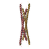
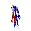
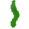
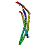
Bicaudal-D uses a parallel, homodimeric coiled coil with heterotypic registry to co-ordinate recruitment of cargos to dynein
ComponentsPROTEIN BICAUDAL D
KeywordsPROTEIN TRANSPORT / CARGO BINDING
Function / homology
Function and homology information


oocyte nucleus migration involved in oocyte dorsal/ventral axis specification / cellular macromolecule localization / microtubule anchoring at microtubule organizing center / germarium-derived egg chamber formation / oocyte microtubule cytoskeleton polarization / germarium-derived oocyte fate determination / positive regulation of synaptic vesicle exocytosis / positive regulation of clathrin-dependent endocytosis / COPI-independent Golgi-to-ER retrograde traffic / protein localization to organelle ...oocyte nucleus migration involved in oocyte dorsal/ventral axis specification / cellular macromolecule localization / microtubule anchoring at microtubule organizing center / germarium-derived egg chamber formation / oocyte microtubule cytoskeleton polarization / germarium-derived oocyte fate determination / positive regulation of synaptic vesicle exocytosis / positive regulation of clathrin-dependent endocytosis / COPI-independent Golgi-to-ER retrograde traffic / protein localization to organelle / oocyte axis specification / RNA transport / clathrin heavy chain binding / intracellular mRNA localization / cytoskeletal anchor activity / dynein complex binding / oogenesis / dynactin binding / regulation of endocytosis / synaptic vesicle endocytosis / mRNA transport / regulation of microtubule cytoskeleton organization / small GTPase binding / presynapse / cytoskeleton / Golgi apparatus / cytoplasm / cytosol
Similarity search - Function
Single alpha-helices involved in coiled-coils or other helix-helix interfaces - #2470 / Bicaudal-D protein / Microtubule-associated protein Bicaudal-D / Single alpha-helices involved in coiled-coils or other helix-helix interfaces / Helix non-globular / Special
Similarity search - Domain/homology
ARGININE / Protein bicaudal D
Similarity search - Component
Biological speciesDROSOPHILA MELANOGASTER (fruit fly)
MethodX-RAY DIFFRACTION / SYNCHROTRON / MAD / Resolution: 2.18 Å
AuthorsLiu, Y. / Salter, H.K. / Holding, A.N. / Johnson, C.M. / Stephens, E. / Lukavsky, P.J. / Walshaw, J. / Bullock, S.L.
CitationJournal: Genes Dev. / Year: 2013
Title: Bicaudal-D Uses a Parallel, Homodimeric Coiled Coil with Heterotypic Registry to Coordinate Recruitment of Cargos to Dynein
Authors: Liu, Y. / Salter, H.K. / Holding, A.N. / Johnson, C.M. / Stephens, E. / Lukavsky, P.J. / Walshaw, J. / Bullock, S.L.
History
DepositionMay 2, 2013Deposition site: PDBE / Processing site: PDBE
Revision 1.0Jun 12, 2013Provider: repository / Type: Initial release
Revision 1.1Jun 26, 2013Group: Database references
Revision 1.2Jul 12, 2017Group: Derived calculations / Category: struct_conn
Item: _struct_conn.pdbx_dist_value / _struct_conn.pdbx_leaving_atom_flag ..._struct_conn.pdbx_dist_value / _struct_conn.pdbx_leaving_atom_flag / _struct_conn.ptnr1_auth_comp_id / _struct_conn.ptnr1_auth_seq_id / _struct_conn.ptnr1_label_atom_id / _struct_conn.ptnr1_label_comp_id / _struct_conn.ptnr1_label_seq_id / _struct_conn.ptnr2_auth_comp_id / _struct_conn.ptnr2_auth_seq_id / _struct_conn.ptnr2_label_atom_id / _struct_conn.ptnr2_label_comp_id / _struct_conn.ptnr2_label_seq_id

-
Structure visualization

Structure viewerMolecule:
MolmilJmol/JSmol

Downloads & links

-
Assembly

Deposited unit
A: PROTEIN BICAUDAL D
B: PROTEIN BICAUDAL D
C: PROTEIN BICAUDAL D
D: PROTEIN BICAUDAL D
hetero molecules


Theoretical massNumber of molelcules
Total (without water)45,1995
Polymers45,0244
Non-polymers1751
Water1,910106
1


  • Idetical with deposited unit
  • defined by author&software
TypeNameSymmetry operationNumber
identity operation1_555x,y,z1
Buried area9780 Å2
ΔGint-90.5 kcal/mol
Surface area21610 Å2
MethodPISA
Unit cell
Length a, b, c (Å)62.199, 62.199, 190.511
Angle α, β, γ (deg.)90.00, 90.00, 120.00
Int Tables number169
Space group name H-MP61

-
Components

#1: Protein
PROTEIN BICAUDAL D


Mass: 11256.005 Da / Num. of mol.: 4 / Fragment: CARGO BINDING DOMAIN, RESIDUES 656-745
Source method: isolated from a genetically manipulated source
Source: (gene. exp.) DROSOPHILA MELANOGASTER (fruit fly) / Description: FLY CDNA / Production host: ESCHERICHIA COLI (E. coli) / References: UniProt: P16568
#2: Chemical ChemComp-ARG / ARGININE


Type: L-peptide linking / Mass: 175.209 Da / Num. of mol.: 1 / Source method: obtained synthetically / Formula: C6H15N4O2
#3: Water ChemComp-HOH / water


Mass: 18.015 Da / Num. of mol.: 106 / Source method: isolated from a natural source / Formula: H2O

-
Experimental details

-
Experiment

ExperimentMethod: X-RAY DIFFRACTION / Number of used crystals: 2

-
Sample preparation

CrystalDensity Matthews: 2.42 Å3/Da / Density % sol: 49 % / Description: NONE
Crystal growpH: 8.5
Details: 0.1 M TRIS PH 8.5, 5 % PEG4000, 10 % GLYCEROL, 30 MM MAGNESIUM CHLORIDE, 30 MM CALCIUM CHLORIDE

-
Data collection

DiffractionMean temperature: 100 K
Diffraction sourceSource: SYNCHROTRON / Site: Diamond / Beamline: I03 / Wavelength: 0.98055
DetectorType: ADSC CCD / Detector: CCD / Date: Mar 10, 2010
RadiationProtocol: MAD / Monochromatic (M) / Laue (L): M / Scattering type: x-ray
Radiation wavelengthWavelength: 0.98055 Å / Relative weight: 1
Reflection twin
Crystal-IDIDOperatorDomain-IDFraction
11H, K, L10.523
11-H-K, K, -L20.477
ReflectionResolution: 2.2→47 Å / Num. obs: 20073 / % possible obs: 100 % / Observed criterion σ(I): 2 / Redundancy: 34 % / Rmerge(I) obs: 0.17 / Net I/σ(I): 18
Reflection shellResolution: 2.18→2.23 Å / Redundancy: 34.7 % / Rmerge(I) obs: 0.17 / Mean I/σ(I) obs: 18 / % possible all: 100

-
Processing

Software
NameVersionClassification
REFMAC5.5.0109refinement
MOSFLMdata reduction
SCALEPACKdata scaling
RefinementMethod to determine structure: MAD
Starting model: NONE

Resolution: 2.18→46.89 Å / Cor.coef. Fo:Fc: 0.933 / Cor.coef. Fo:Fc free: 0.915 / SU B: 2.598 / SU ML: 0.07 / Cross valid method: THROUGHOUT / ESU R: 0.051 / ESU R Free: 0.04 / Stereochemistry target values: MAXIMUM LIKELIHOOD / Details: HYDROGENS HAVE BEEN ADDED IN THE RIDING POSITIONS.
RfactorNum. reflection% reflectionSelection details
Rfree0.23518 1746 8 %RANDOM
Rwork0.20367 ---
obs0.2061 20073 99.86 %-
Solvent computationIon probe radii: 0.8 Å / Shrinkage radii: 0.8 Å / VDW probe radii: 1.4 Å / Solvent model: MASK
Displacement parametersBiso mean: 36.344 Å2
Baniso -1Baniso -2Baniso -3
1-19.37 Å20 Å20 Å2
2--19.37 Å20 Å2
3----38.75 Å2
Refinement stepCycle: LAST / Resolution: 2.18→46.89 Å
ProteinNucleic acidLigandSolventTotal
Num. atoms2568 0 4 106 2678
Refine LS restraints
Refine-IDTypeDev idealDev ideal targetNumber
X-RAY DIFFRACTIONr_bond_refined_d0.0270.0222580
X-RAY DIFFRACTIONr_bond_other_d
X-RAY DIFFRACTIONr_angle_refined_deg2.4261.9913464
X-RAY DIFFRACTIONr_angle_other_deg
X-RAY DIFFRACTIONr_dihedral_angle_1_deg6.7945324
X-RAY DIFFRACTIONr_dihedral_angle_2_deg40.21225124
X-RAY DIFFRACTIONr_dihedral_angle_3_deg22.40315525
X-RAY DIFFRACTIONr_dihedral_angle_4_deg18.3081525
X-RAY DIFFRACTIONr_chiral_restr0.1690.2424
X-RAY DIFFRACTIONr_gen_planes_refined0.0110.021857
X-RAY DIFFRACTIONr_gen_planes_other
X-RAY DIFFRACTIONr_nbd_refined
X-RAY DIFFRACTIONr_nbd_other
X-RAY DIFFRACTIONr_nbtor_refined
X-RAY DIFFRACTIONr_nbtor_other
X-RAY DIFFRACTIONr_xyhbond_nbd_refined
X-RAY DIFFRACTIONr_xyhbond_nbd_other
X-RAY DIFFRACTIONr_metal_ion_refined
X-RAY DIFFRACTIONr_metal_ion_other
X-RAY DIFFRACTIONr_symmetry_vdw_refined
X-RAY DIFFRACTIONr_symmetry_vdw_other
X-RAY DIFFRACTIONr_symmetry_hbond_refined
X-RAY DIFFRACTIONr_symmetry_hbond_other
X-RAY DIFFRACTIONr_symmetry_metal_ion_refined
X-RAY DIFFRACTIONr_symmetry_metal_ion_other
X-RAY DIFFRACTIONr_mcbond_it1.3661.51634
X-RAY DIFFRACTIONr_mcbond_other
X-RAY DIFFRACTIONr_mcangle_it2.2122596
X-RAY DIFFRACTIONr_mcangle_other
X-RAY DIFFRACTIONr_scbond_it4.563946
X-RAY DIFFRACTIONr_scbond_other
X-RAY DIFFRACTIONr_scangle_it6.794.5868
X-RAY DIFFRACTIONr_scangle_other
X-RAY DIFFRACTIONr_long_range_B_refined
X-RAY DIFFRACTIONr_long_range_B_other
X-RAY DIFFRACTIONr_rigid_bond_restr
X-RAY DIFFRACTIONr_sphericity_free
X-RAY DIFFRACTIONr_sphericity_bonded
LS refinement shellResolution: 2.175→2.231 Å / Total num. of bins used: 20
RfactorNum. reflection% reflection
Rfree0.238 111 -
Rwork0.192 1492 -
obs--100 %

+
About Yorodumi

-
News

-
Feb 9, 2022. New format data for meta-information of EMDB entries

New format data for meta-information of EMDB entries

  • Version 3 of the EMDB header file is now the official format.
  • The previous official version 1.9 will be removed from the archive.

Related info.:EMDB header

External links:wwPDB to switch to version 3 of the EMDB data model

-
Aug 12, 2020. Covid-19 info

Covid-19 info

URL: https://pdbjlvh1.pdbj.org/emnavi/covid19.php

New page: Covid-19 featured information page in EM Navigator.

Related info.:Covid-19 info / Mar 5, 2020. Novel coronavirus structure data

+
Mar 5, 2020. Novel coronavirus structure data

Novel coronavirus structure data

Related info.:Yorodumi Speices / Aug 12, 2020. Covid-19 info

External links:COVID-19 featured content - PDBj / Molecule of the Month (242):Coronavirus Proteases

+
Jan 31, 2019. EMDB accession codes are about to change! (news from PDBe EMDB page)

EMDB accession codes are about to change! (news from PDBe EMDB page)

  • The allocation of 4 digits for EMDB accession codes will soon come to an end. Whilst these codes will remain in use, new EMDB accession codes will include an additional digit and will expand incrementally as the available range of codes is exhausted. The current 4-digit format prefixed with “EMD-” (i.e. EMD-XXXX) will advance to a 5-digit format (i.e. EMD-XXXXX), and so on. It is currently estimated that the 4-digit codes will be depleted around Spring 2019, at which point the 5-digit format will come into force.
  • The EM Navigator/Yorodumi systems omit the EMD- prefix.

Related info.:Q: What is EMD? / ID/Accession-code notation in Yorodumi/EM Navigator

External links:EMDB Accession Codes are Changing Soon! / Contact to PDBj

+
Jul 12, 2017. Major update of PDB

Major update of PDB

  • wwPDB released updated PDB data conforming to the new PDBx/mmCIF dictionary.
  • This is a major update changing the version number from 4 to 5, and with Remediation, in which all the entries are updated.
  • In this update, many items about electron microscopy experimental information are reorganized (e.g. em_software).
  • Now, EM Navigator and Yorodumi are based on the updated data.

External links:wwPDB Remediation / Enriched Model Files Conforming to OneDep Data Standards Now Available in the PDB FTP Archive

-
Yorodumi

Thousand views of thousand structures

  • Yorodumi is a browser for structure data from EMDB, PDB, SASBDB, etc.
  • This page is also the successor to EM Navigator detail page, and also detail information page/front-end page for Omokage search.
  • The word "yorodu" (or yorozu) is an old Japanese word meaning "ten thousand". "mi" (miru) is to see.

Related info.:EMDB / PDB / SASBDB / Comparison of 3 databanks / Yorodumi Search / Aug 31, 2016. New EM Navigator & Yorodumi / Yorodumi Papers / Jmol/JSmol / Function and homology information / Changes in new EM Navigator and Yorodumi

Read more