[English] 日本語
Yorodumi
- PDB-4bk0: Crystal structure of the KIX domain of human RECQL5 (domain-swapp... -

+
Open data


ID or keywords:

Loading...

-
Basic information

Entry
Database: PDB / ID: 4bk0
TitleCrystal structure of the KIX domain of human RECQL5 (domain-swapped dimer)
ComponentsATP-DEPENDENT DNA HELICASE Q5
KeywordsTRANSCRIPTION / DNA HELICASE / TRANSCRIPTIONAL REPRESSION
Function / homology
Function and homology information


mitotic DNA-templated DNA replication / chromosome separation / cellular response to camptothecin / four-way junction helicase activity / replication-born double-strand break repair via sister chromatid exchange / DNA 3'-5' helicase / transcription preinitiation complex / DNA metabolic process / RNA polymerase II complex binding / negative regulation of transcription elongation by RNA polymerase II ...mitotic DNA-templated DNA replication / chromosome separation / cellular response to camptothecin / four-way junction helicase activity / replication-born double-strand break repair via sister chromatid exchange / DNA 3'-5' helicase / transcription preinitiation complex / DNA metabolic process / RNA polymerase II complex binding / negative regulation of transcription elongation by RNA polymerase II / DNA unwinding involved in DNA replication / 3'-5' DNA helicase activity / negative regulation of double-strand break repair via homologous recombination / DNA helicase activity / isomerase activity / helicase activity / replication fork / double-strand break repair via homologous recombination / cellular response to xenobiotic stimulus / chromosome / mitotic cell cycle / DNA replication / cell division / DNA repair / ATP hydrolysis activity / DNA binding / nucleoplasm / ATP binding / identical protein binding / nucleus / metal ion binding / cytoplasm / cytosol
Similarity search - Function
Single alpha-helices involved in coiled-coils or other helix-helix interfaces - #2460 / Single alpha-helices involved in coiled-coils or other helix-helix interfaces - #3140 / RecQ helicase-like 5 / RecQ helicase protein-like 5 (RecQ5) / ATP-dependent DNA helicase RecQ, zinc-binding domain / RecQ zinc-binding / DNA helicase, ATP-dependent, RecQ type / DNA/RNA helicase, ATP-dependent, DEAH-box type, conserved site / DEAH-box subfamily ATP-dependent helicases signature. / Single alpha-helices involved in coiled-coils or other helix-helix interfaces ...Single alpha-helices involved in coiled-coils or other helix-helix interfaces - #2460 / Single alpha-helices involved in coiled-coils or other helix-helix interfaces - #3140 / RecQ helicase-like 5 / RecQ helicase protein-like 5 (RecQ5) / ATP-dependent DNA helicase RecQ, zinc-binding domain / RecQ zinc-binding / DNA helicase, ATP-dependent, RecQ type / DNA/RNA helicase, ATP-dependent, DEAH-box type, conserved site / DEAH-box subfamily ATP-dependent helicases signature. / Single alpha-helices involved in coiled-coils or other helix-helix interfaces / DEAD/DEAH box helicase / DEAD/DEAH box helicase domain / Helix non-globular / Helicase conserved C-terminal domain / Special / helicase superfamily c-terminal domain / Superfamilies 1 and 2 helicase C-terminal domain profile. / Superfamilies 1 and 2 helicase ATP-binding type-1 domain profile. / DEAD-like helicases superfamily / Helicase, C-terminal / Helicase superfamily 1/2, ATP-binding domain / P-loop containing nucleoside triphosphate hydrolase
Similarity search - Domain/homology
DI(HYDROXYETHYL)ETHER / ATP-dependent DNA helicase Q5
Similarity search - Component
Biological speciesHOMO SAPIENS (human)
MethodX-RAY DIFFRACTION / SYNCHROTRON / SAD / Resolution: 1.9 Å
AuthorsKassube, S.A. / Jinek, M. / Fang, J. / Tsutakawa, S. / Nogales, E.
Citation
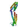
Journal: Nat Struct Mol Biol / Year: 2013
Title: Structural mimicry in transcription regulation of human RNA polymerase II by the DNA helicase RECQL5.
Authors: Susanne A Kassube / Martin Jinek / Jie Fang / Susan Tsutakawa / Eva Nogales /
Abstract: RECQL5 is a member of the highly conserved RecQ family of DNA helicases involved in DNA repair. RECQL5 interacts with RNA polymerase II (Pol II) and inhibits transcription of protein-encoding genes ...RECQL5 is a member of the highly conserved RecQ family of DNA helicases involved in DNA repair. RECQL5 interacts with RNA polymerase II (Pol II) and inhibits transcription of protein-encoding genes by an unknown mechanism. We show that RECQL5 contacts the Rpb1 jaw domain of Pol II at a site that overlaps with the binding site for the transcription elongation factor TFIIS. Our cryo-EM structure of elongating Pol II arrested in complex with RECQL5 shows that the RECQL5 helicase domain is positioned to sterically block elongation. The crystal structure of the RECQL5 KIX domain reveals similarities with TFIIS, and binding of RECQL5 to Pol II interferes with the ability of TFIIS to promote transcriptional read-through in vitro. Together, our findings reveal a dual mode of transcriptional repression by RECQL5 that includes structural mimicry of the Pol II-TFIIS interaction.
History
DepositionApr 21, 2013Deposition site: PDBE / Processing site: PDBE
Revision 1.0Jun 12, 2013Provider: repository / Type: Initial release
Revision 1.1Jun 19, 2013Group: Atomic model / Database references
Revision 1.2Jul 17, 2013Group: Database references
Revision 1.3Nov 13, 2019Group: Data collection / Database references / Other / Category: pdbx_database_related / pdbx_database_status
Item: _pdbx_database_related.content_type / _pdbx_database_status.status_code_sf
Revision 1.4May 8, 2024Group: Data collection / Database references / Derived calculations
Category: chem_comp_atom / chem_comp_bond ...chem_comp_atom / chem_comp_bond / database_2 / struct_site
Item: _database_2.pdbx_DOI / _database_2.pdbx_database_accession ..._database_2.pdbx_DOI / _database_2.pdbx_database_accession / _struct_site.pdbx_auth_asym_id / _struct_site.pdbx_auth_comp_id / _struct_site.pdbx_auth_seq_id

-
Structure visualization

Structure viewerMolecule:
MolmilJmol/JSmol

Downloads & links

-
Assembly

Deposited unit
A: ATP-DEPENDENT DNA HELICASE Q5
B: ATP-DEPENDENT DNA HELICASE Q5
hetero molecules


Theoretical massNumber of molelcules
Total (without water)25,2534
Polymers25,0412
Non-polymers2122
Water1,69394
1


  • Idetical with deposited unit
  • defined by software
TypeNameSymmetry operationNumber
identity operation1_555x,y,z1
Buried area5640 Å2
ΔGint-28 kcal/mol
Surface area12070 Å2
MethodPISA
Unit cell
Length a, b, c (Å)50.670, 61.210, 81.650
Angle α, β, γ (deg.)90.00, 90.00, 90.00
Int Tables number19
Space group name H-MP212121

-
Components

#1: Protein ATP-DEPENDENT DNA HELICASE Q5 / DNA HELICASE / RECQ-LIKE TYPE 5 / RECQ5 / RECQ PROTEIN-LIKE 5 / RECQL5


Mass: 12520.251 Da / Num. of mol.: 2 / Fragment: KIX DOMAIN, RESIDUES 515-620
Source method: isolated from a genetically manipulated source
Source: (gene. exp.) HOMO SAPIENS (human) / Production host: ESCHERICHIA COLI (E. coli) / Strain (production host): BL21 / Variant (production host): RIL / References: UniProt: O94762, DNA helicase
#2: Chemical ChemComp-PEG / DI(HYDROXYETHYL)ETHER


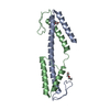
Mass: 106.120 Da / Num. of mol.: 2 / Source method: obtained synthetically / Formula: C4H10O3
#3: Water ChemComp-HOH / water


Mass: 18.015 Da / Num. of mol.: 94 / Source method: isolated from a natural source / Formula: H2O
Sequence detailsGLPGS SEQUENCE AT N TERMINUS IS DERIVED FROM THE EXPRESSION VECTOR

-
Experimental details

-
Experiment

ExperimentMethod: X-RAY DIFFRACTION / Number of used crystals: 1

-
Sample preparation

CrystalDensity Matthews: 2.53 Å3/Da / Density % sol: 51.5 % / Description: NONE
Crystal growpH: 6.2
Details: 0.2 M NACL, 0.1 M SODIUM/POTASSIUM PHOSPHATE PH 6.2, 50% PEG 200

-
Data collection

DiffractionMean temperature: 100 K
Diffraction sourceSource: SYNCHROTRON / Site: ALS / Beamline: 8.3.1 / Wavelength: 1.11589
DetectorType: ADSC CCD / Detector: CCD / Date: Oct 9, 2010
RadiationProtocol: SINGLE WAVELENGTH / Monochromatic (M) / Laue (L): M / Scattering type: x-ray
Radiation wavelengthWavelength: 1.11589 Å / Relative weight: 1
ReflectionResolution: 1.9→49 Å / Num. obs: 20613 / % possible obs: 100 % / Observed criterion σ(I): 2 / Redundancy: 7 % / Biso Wilson estimate: 31.7 Å2 / Rmerge(I) obs: 0.04 / Net I/σ(I): 22.8
Reflection shellResolution: 1.9→1.95 Å / Redundancy: 7.1 % / Rmerge(I) obs: 0.52 / Mean I/σ(I) obs: 3.6 / % possible all: 100

-
Processing

Software
NameVersionClassification
PHENIX(PHENIX.REFINE)refinement
XDSdata reduction
XSCALEdata scaling
autoSHARPphasing
RefinementMethod to determine structure: SAD
Starting model: NONE

Resolution: 1.9→48.976 Å / SU ML: 0.21 / σ(F): 2 / Phase error: 20.72 / Stereochemistry target values: ML
RfactorNum. reflection% reflection
Rfree0.2336 1054 5.1 %
Rwork0.2014 --
obs0.203 20613 99.9 %
Solvent computationShrinkage radii: 0.86 Å / VDW probe radii: 1.1 Å / Solvent model: FLAT BULK SOLVENT MODEL / Bsol: 48.041 Å2 / ksol: 0.371 e/Å3
Displacement parametersBiso mean: 48.44 Å2
Baniso -1Baniso -2Baniso -3
1--7.933 Å20 Å20 Å2
2--11.9549 Å20 Å2
3----4.0219 Å2
Refinement stepCycle: LAST / Resolution: 1.9→48.976 Å
ProteinNucleic acidLigandSolventTotal
Num. atoms1504 0 14 94 1612
Refine LS restraints
Refine-IDTypeDev idealNumber
X-RAY DIFFRACTIONf_bond_d0.0091542
X-RAY DIFFRACTIONf_angle_d1.0432071
X-RAY DIFFRACTIONf_dihedral_angle_d13.781616
X-RAY DIFFRACTIONf_chiral_restr0.052233
X-RAY DIFFRACTIONf_plane_restr0.006274
LS refinement shell
Resolution (Å)Rfactor RfreeNum. reflection RfreeRfactor RworkNum. reflection RworkRefine-ID% reflection obs (%)
1.9-1.98650.22731260.23332394X-RAY DIFFRACTION100
1.9865-2.09120.23611270.19892396X-RAY DIFFRACTION100
2.0912-2.22220.19571260.19272406X-RAY DIFFRACTION100
2.2222-2.39380.26551480.18472404X-RAY DIFFRACTION100
2.3938-2.63470.23131380.19432431X-RAY DIFFRACTION100
2.6347-3.01590.24961400.19722441X-RAY DIFFRACTION100
3.0159-3.79950.24311020.21122516X-RAY DIFFRACTION100
3.7995-48.99220.22211470.20072571X-RAY DIFFRACTION100
Refinement TLS params.

Method: refined / Refine-ID: X-RAY DIFFRACTION

IDL112)L122)L132)L222)L232)L332)S11 (Å °)S12 (Å °)S13 (Å °)S21 (Å °)S22 (Å °)S23 (Å °)S31 (Å °)S32 (Å °)S33 (Å °)T112)T122)T132)T222)T232)T332)Origin x (Å)Origin y (Å)Origin z (Å)
17.05430.63770.6481.86761.60054.2246-0.05650.68360.6063-0.25720.06070.1504-0.54340.11270.00020.2291-0.01690.01930.27790.02950.259-1.987353.1082-5.9587
25.3304-0.18797.3337-1.23420.08146.82130.1008-0.5008-0.4260.3820.1117-0.08040.1268-0.3086-0.22470.46740.0311-0.09190.47070.00660.343414.849451.352722.4886
35.62390.04921.04746.6086-0.29294.61440.21910.9017-0.6689-2.0851-0.309-1.44580.85921.50520.08380.68040.47920.18950.397-0.00490.439639.713752.155934.2388
47.1122-0.8792-1.63136.9755-1.53784.6445-0.1719-0.0136-0.4935-0.18030.042-0.02690.8374-0.0403-0.0520.33790.0266-0.04180.1690.02520.266228.651355.802440.708
50.06140.7313-0.65818.204-7.39846.6582-1.48330.2202-0.00012.87920.6152-1.6704-1.85022.14791.27360.61410.1323-0.31640.7337-0.21880.658521.625649.870313.5796
65.13351.14281.78863.3581.28055.40250.2349-0.6349-0.6970.56680.1137-0.15310.53560.0103-0.30610.25720.03530.00190.27150.08040.27942.30744.95749.6607
Refinement TLS group
IDRefine-IDRefine TLS-IDSelection details
1X-RAY DIFFRACTION1CHAIN A AND (RESSEQ 523:546)
2X-RAY DIFFRACTION2CHAIN A AND (RESSEQ 547:589)
3X-RAY DIFFRACTION3CHAIN A AND (RESSEQ 590:614)
4X-RAY DIFFRACTION4CHAIN B AND (RESSEQ 524:568)
5X-RAY DIFFRACTION5CHAIN B AND (RESSEQ 569:575)
6X-RAY DIFFRACTION6CHAIN B AND (RESSEQ 576:620)

+
About Yorodumi

-
News

-
Feb 9, 2022. New format data for meta-information of EMDB entries

New format data for meta-information of EMDB entries

  • Version 3 of the EMDB header file is now the official format.
  • The previous official version 1.9 will be removed from the archive.

Related info.:EMDB header

External links:wwPDB to switch to version 3 of the EMDB data model

-
Aug 12, 2020. Covid-19 info

Covid-19 info

URL: https://pdbjlvh1.pdbj.org/emnavi/covid19.php

New page: Covid-19 featured information page in EM Navigator.

Related info.:Covid-19 info / Mar 5, 2020. Novel coronavirus structure data

+
Mar 5, 2020. Novel coronavirus structure data

Novel coronavirus structure data

Related info.:Yorodumi Speices / Aug 12, 2020. Covid-19 info

External links:COVID-19 featured content - PDBj / Molecule of the Month (242):Coronavirus Proteases

+
Jan 31, 2019. EMDB accession codes are about to change! (news from PDBe EMDB page)

EMDB accession codes are about to change! (news from PDBe EMDB page)

  • The allocation of 4 digits for EMDB accession codes will soon come to an end. Whilst these codes will remain in use, new EMDB accession codes will include an additional digit and will expand incrementally as the available range of codes is exhausted. The current 4-digit format prefixed with “EMD-” (i.e. EMD-XXXX) will advance to a 5-digit format (i.e. EMD-XXXXX), and so on. It is currently estimated that the 4-digit codes will be depleted around Spring 2019, at which point the 5-digit format will come into force.
  • The EM Navigator/Yorodumi systems omit the EMD- prefix.

Related info.:Q: What is EMD? / ID/Accession-code notation in Yorodumi/EM Navigator

External links:EMDB Accession Codes are Changing Soon! / Contact to PDBj

+
Jul 12, 2017. Major update of PDB

Major update of PDB

  • wwPDB released updated PDB data conforming to the new PDBx/mmCIF dictionary.
  • This is a major update changing the version number from 4 to 5, and with Remediation, in which all the entries are updated.
  • In this update, many items about electron microscopy experimental information are reorganized (e.g. em_software).
  • Now, EM Navigator and Yorodumi are based on the updated data.

External links:wwPDB Remediation / Enriched Model Files Conforming to OneDep Data Standards Now Available in the PDB FTP Archive

-
Yorodumi

Thousand views of thousand structures

  • Yorodumi is a browser for structure data from EMDB, PDB, SASBDB, etc.
  • This page is also the successor to EM Navigator detail page, and also detail information page/front-end page for Omokage search.
  • The word "yorodu" (or yorozu) is an old Japanese word meaning "ten thousand". "mi" (miru) is to see.

Related info.:EMDB / PDB / SASBDB / Comparison of 3 databanks / Yorodumi Search / Aug 31, 2016. New EM Navigator & Yorodumi / Yorodumi Papers / Jmol/JSmol / Function and homology information / Changes in new EM Navigator and Yorodumi

Read more