 Movie
Movie Controller


+ Open data

- Basic information
| Entry | Database: PDB / ID: 4lpv | ||||||
|---|---|---|---|---|---|---|---|
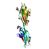
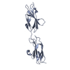
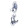
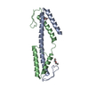
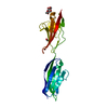
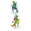
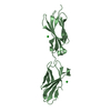
| Title | Crystal structure of TENCON variant P41BR3-42 | ||||||
 Components Components | TENCON variant P41BR3-42 | ||||||
 Keywords Keywords | DE NOVO PROTEIN / fibronectin type III fold / alternate scaffold | ||||||
| Function / homology | Immunoglobulins / Immunoglobulin-like / Sandwich / Mainly Beta Function and homology information Function and homology information | ||||||
| Biological species | artificial gene (others) | ||||||
| Method |  X-RAY DIFFRACTION / MOLECULAR REPLACEMENT / Resolution: 1.8 Å X-RAY DIFFRACTION / MOLECULAR REPLACEMENT / Resolution: 1.8 Å | ||||||
 Authors Authors | Teplyakov, A. / Obmolova, G. / Luo, J. / Gilliland, G.L. | ||||||
 Citation Citation | Journal: Proteins / Year: 2014 Title: C-terminal beta-strand swapping in a consensus-derived fibronectin Type III scaffold. Authors: Teplyakov, A. / Obmolova, G. / Malia, T.J. / Luo, J. / Jacobs, S.A. / Chan, W. / Domingo, D. / Baker, A. / O'Neil, K.T. / Gilliland, G.L. | ||||||
| History |
|
- Structure visualization
| Structure viewer | Molecule: MolmilJmol/JSmol |
|---|
- Downloads & links
-Download
| PDBx/mmCIF format | 4lpv.cif.gz | 59.2 KB | Display | PDBx/mmCIF format |
|---|---|---|---|---|
| PDB format | pdb4lpv.ent.gz | 42.4 KB | Display | PDB format |
| PDBx/mmJSON format | 4lpv.json.gz | Tree view | PDBx/mmJSON format | |
| Others |  Other downloads Other downloads |
-Validation report
| Arichive directory | https://data.pdbj.org/pub/pdb/validation_reports/lp/4lpvftp://data.pdbj.org/pub/pdb/validation_reports/lp/4lpv | HTTPS FTP |
|---|
-Related structure data
| Related structure data |  4lptC  4lpuC  4lpwC  4lpxC  4lpyC  3tesS C: citing same article ( S: Starting model for refinement |
|---|---|
| Similar structure data |


-Links
 PDBj
PDBj
- Assembly
| Deposited unit | 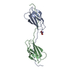
| ||||||||||||||||||
|---|---|---|---|---|---|---|---|---|---|---|---|---|---|---|---|---|---|---|---|
| 1 |
| ||||||||||||||||||
| 2 | 
| ||||||||||||||||||
| Unit cell |
| ||||||||||||||||||
| Components on special symmetry positions |
|
-Components
| #1: Protein | Mass: 10949.251 Da / Num. of mol.: 2 Source method: isolated from a genetically manipulated source Source: (gene. exp.) artificial gene (others) / Production host:  #2: Chemical | ChemComp-TRS / |   Mass: 122.143 Da / Num. of mol.: 1 / Source method: obtained synthetically / Formula: C4H12NO3 / Comment: pH buffer*YM Mass: 122.143 Da / Num. of mol.: 1 / Source method: obtained synthetically / Formula: C4H12NO3 / Comment: pH buffer*YM#3: Water | ChemComp-HOH / |  Mass: 18.015 Da / Num. of mol.: 364 / Source method: isolated from a natural source / Formula: H2O Mass: 18.015 Da / Num. of mol.: 364 / Source method: isolated from a natural source / Formula: H2O |
|---|
-Experimental details
-Experiment
| Experiment | Method: X-RAY DIFFRACTION / Number of used crystals: 1 |
|---|
- Sample preparation
| Crystal | Density Matthews: 5.74 Å3/Da / Density % sol: 78.58 % |
|---|---|
| Crystal grow | Temperature: 293 K / Method: vapor diffusion, sitting drop / pH: 8.5 Details: 0.1 M Tris, pH 8.5, 1.35 M ammonium sulfate, 5% PEG400, VAPOR DIFFUSION, SITTING DROP, temperature 293K |
-Data collection
| Diffraction | Mean temperature: 95 K |
|---|---|
| Diffraction source | Source: ROTATING ANODE / Type: RIGAKU MICROMAX-007 HF / Wavelength: 1.5418 / Wavelength: 1.5418 Å |
| Detector | Type: RIGAKU SATURN 944 / Detector: CCD / Date: Nov 9, 2009 / Details: VARIMAX HF |
| Radiation | Protocol: SINGLE WAVELENGTH / Monochromatic (M) / Laue (L): M / Scattering type: x-ray |
| Radiation wavelength | Wavelength: 1.5418 Å / Relative weight: 1 |
| Reflection | Resolution: 1.8→49 Å / Num. all: 42629 / Num. obs: 42629 / % possible obs: 94.8 % / Observed criterion σ(F): 0 / Observed criterion σ(I): -3 / Redundancy: 4.9 % / Rmerge(I) obs: 0.043 |
| Reflection shell | Resolution: 1.8→1.85 Å / Redundancy: 2.1 % / Rmerge(I) obs: 0.369 / % possible all: 62.8 |
- Processing
| Software |
| |||||||||||||||||||||||||||||||||||||||||||||||||||||||||||||||||||||||||||||||||||||||||||||||||||||||||
|---|---|---|---|---|---|---|---|---|---|---|---|---|---|---|---|---|---|---|---|---|---|---|---|---|---|---|---|---|---|---|---|---|---|---|---|---|---|---|---|---|---|---|---|---|---|---|---|---|---|---|---|---|---|---|---|---|---|---|---|---|---|---|---|---|---|---|---|---|---|---|---|---|---|---|---|---|---|---|---|---|---|---|---|---|---|---|---|---|---|---|---|---|---|---|---|---|---|---|---|---|---|---|---|---|---|---|
| Refinement | Method to determine structure: MOLECULAR REPLACEMENT Starting model: PDB ENTRY 3TES Resolution: 1.8→31.1 Å / σ(F): 2 / Phase error: 21.16 / Stereochemistry target values: Engh & Huber
| |||||||||||||||||||||||||||||||||||||||||||||||||||||||||||||||||||||||||||||||||||||||||||||||||||||||||
| Solvent computation | Shrinkage radii: 0.47 Å / VDW probe radii: 0.8 Å / Solvent model: FLAT BULK SOLVENT MODEL / Bsol: 49.965 Å2 / ksol: 0.427 e/Å3 | |||||||||||||||||||||||||||||||||||||||||||||||||||||||||||||||||||||||||||||||||||||||||||||||||||||||||
| Displacement parameters |
| |||||||||||||||||||||||||||||||||||||||||||||||||||||||||||||||||||||||||||||||||||||||||||||||||||||||||
| Refinement step | Cycle: LAST / Resolution: 1.8→31.1 Å
| |||||||||||||||||||||||||||||||||||||||||||||||||||||||||||||||||||||||||||||||||||||||||||||||||||||||||
| Refine LS restraints |
| |||||||||||||||||||||||||||||||||||||||||||||||||||||||||||||||||||||||||||||||||||||||||||||||||||||||||
| LS refinement shell |
|
