 Movie
Movie Controller

[English] 日本語
 Yorodumi
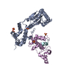
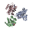
- PDB-4bj5: Crystal structure of Rif2 in complex with the C-terminal domain o... -
+ Open data

- Basic information
| Entry | Database: PDB / ID: 4bj5 | ||||||
|---|---|---|---|---|---|---|---|
| Title | Crystal structure of Rif2 in complex with the C-terminal domain of Rap1 (Rap1-RCT) | ||||||
 Components Components |
| ||||||
 Keywords Keywords | TRANSCRIPTION / GENOME STABILITY / TELOMERE ASSOCIATED PROTEINS / AAA+ FOLD | ||||||
| Function / homology |  Function and homology information Function and homology informationpositive regulation of ribosomal protein gene transcription by RNA polymerase II / G-quadruplex DNA formation / telomeric G-quadruplex DNA binding / protection from non-homologous end joining at telomere / establishment of protein localization to telomere / establishment of protein localization to chromatin / telomere maintenance via telomere lengthening / shelterin complex / double-stranded telomeric DNA binding / G-quadruplex DNA binding ...positive regulation of ribosomal protein gene transcription by RNA polymerase II / G-quadruplex DNA formation / telomeric G-quadruplex DNA binding / protection from non-homologous end joining at telomere / establishment of protein localization to telomere / establishment of protein localization to chromatin / telomere maintenance via telomere lengthening / shelterin complex / double-stranded telomeric DNA binding / G-quadruplex DNA binding / telomere capping / silent mating-type cassette heterochromatin formation / regulation of glycolytic process / DNA binding, bending / nucleosomal DNA binding / nuclear chromosome / telomeric repeat DNA binding / TFIID-class transcription factor complex binding / subtelomeric heterochromatin formation / telomere maintenance via telomerase / cis-regulatory region sequence-specific DNA binding / TBP-class protein binding / telomere maintenance / protein-DNA complex / transcription regulator complex / histone binding / sequence-specific DNA binding / RNA polymerase II-specific DNA-binding transcription factor binding / DNA-binding transcription factor activity, RNA polymerase II-specific / chromosome, telomeric region / DNA-binding transcription factor activity / negative regulation of transcription by RNA polymerase II / positive regulation of transcription by RNA polymerase II / nucleus / cytosol Similarity search - Function | ||||||
| Biological species |  | ||||||
| Method |  X-RAY DIFFRACTION / SYNCHROTRON / MOLECULAR REPLACEMENT / Resolution: 3.29 Å X-RAY DIFFRACTION / SYNCHROTRON / MOLECULAR REPLACEMENT / Resolution: 3.29 Å | ||||||
 Authors Authors | Shi, T. / Bunker, R.D. / Gut, H. / Scrima, A. / Thoma, N.H. | ||||||
 Citation Citation | Journal: Cell(Cambridge,Mass.) / Year: 2013 Title: Rif1 and Rif2 Shape Telomere Funcation and Architecture Through Multivalent RAP1 Interactions Authors: Shi, T. / Bunker, R.D. / Mattarocci, S. / Ribeyre, C. / Faty, M. / Gut, H. / Scrima, A. / Rass, U. / Rubin, S.M. / Shore, D. / Thoma, N.H. | ||||||
| History |
|
- Structure visualization
| Structure viewer | Molecule: MolmilJmol/JSmol |
|---|
- Downloads & links
-Download
| PDBx/mmCIF format | 4bj5.cif.gz | 379.1 KB | Display | PDBx/mmCIF format |
|---|---|---|---|---|
| PDB format | pdb4bj5.ent.gz | 311.7 KB | Display | PDB format |
| PDBx/mmJSON format | 4bj5.json.gz | Tree view | PDBx/mmJSON format | |
| Others |  Other downloads Other downloads |
-Validation report
| Arichive directory | https://data.pdbj.org/pub/pdb/validation_reports/bj/4bj5ftp://data.pdbj.org/pub/pdb/validation_reports/bj/4bj5 | HTTPS FTP |
|---|
-Related structure data
| Related structure data | 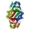 4bj1SC 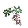 4bj6C 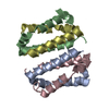 4bjsC 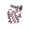 4bjtC 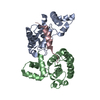 3owtS S: Starting model for refinement C: citing same article ( |
|---|---|
| Similar structure data |


-Links
 PDBj
PDBj


- Assembly
| Deposited unit | 
| ||||||||
|---|---|---|---|---|---|---|---|---|---|
| 1 | 
| ||||||||
| 2 | 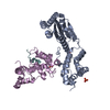
| ||||||||
| Unit cell |
|
-Components
| #1: Protein | Mass: 46036.207 Da / Num. of mol.: 2 Source method: isolated from a genetically manipulated source Source: (gene. exp.) Strain: S288C / Plasmid: PAD DERIVED / Cell line (production host): High Five / Production host:  TRICHOPLUSIA NI (cabbage looper) / References: UniProt: Q06208 TRICHOPLUSIA NI (cabbage looper) / References: UniProt: Q06208#2: Protein | Mass: 23248.975 Da / Num. of mol.: 2 / Fragment: C-TERMINAL DOMAIN, RESIDUES 627-827 Source method: isolated from a genetically manipulated source Source: (gene. exp.) Strain: S288C / Plasmid: PAD DERIVED / Cell line (production host): High Five / Production host: TRICHOPLUSIA NI (cabbage looper) / References: UniProt: P11938#3: Protein/peptide | Mass: 1543.956 Da / Num. of mol.: 2 / Fragment: RESIDUES 36-48 / Source method: obtained synthetically / Source: (synth.) #4: Chemical | ChemComp-SO4 /   Mass: 96.063 Da / Num. of mol.: 5 / Source method: obtained synthetically / Formula: SO4 Mass: 96.063 Da / Num. of mol.: 5 / Source method: obtained synthetically / Formula: SO4 |
|---|
-Experimental details
-Experiment
| Experiment | Method: X-RAY DIFFRACTION / Number of used crystals: 1 |
|---|
- Sample preparation
| Crystal | Density Matthews: 4.1 Å3/Da / Density % sol: 70 % / Description: NONE |
|---|---|
| Crystal grow | Details: 22-25% PEG 6000, 100 MM TRIS/HCL PH 8.0, 500 MM LI2SO4 |
-Data collection
| Diffraction | Mean temperature: 100 K |
|---|---|
| Diffraction source | Source: SYNCHROTRON / Site: SLS  / Beamline: X10SA / Wavelength: 1 / Beamline: X10SA / Wavelength: 1 |
| Detector | Type: DECTRIS PILATUS 6M / Detector: PIXEL / Date: Jul 30, 2010 / Details: MIRRORS |
| Radiation | Monochromator: DOUBLE-CRYSTAL SI(111) MONOCHROMATOR / Protocol: SINGLE WAVELENGTH / Monochromatic (M) / Laue (L): M / Scattering type: x-ray |
| Radiation wavelength | Wavelength: 1 Å / Relative weight: 1 |
| Reflection | Resolution: 3.29→68.5 Å / Num. obs: 27170 / % possible obs: 99.7 % / Observed criterion σ(I): -3 / Redundancy: 3.6 % / Biso Wilson estimate: 119.43 Å2 / Rmerge(I) obs: 0.06 / Net I/σ(I): 14.3 |
| Reflection shell | Resolution: 3.29→3.3 Å / Redundancy: 3.6 % / Rmerge(I) obs: 0.56 / Mean I/σ(I) obs: 2.4 / % possible all: 100 |
- Processing
| Software |
| |||||||||||||||||||||||||||||||||||||||||||||||||||||||||||||||||||||||||||||||||||||||||||||||||||||||||||||||||||||||||||||
|---|---|---|---|---|---|---|---|---|---|---|---|---|---|---|---|---|---|---|---|---|---|---|---|---|---|---|---|---|---|---|---|---|---|---|---|---|---|---|---|---|---|---|---|---|---|---|---|---|---|---|---|---|---|---|---|---|---|---|---|---|---|---|---|---|---|---|---|---|---|---|---|---|---|---|---|---|---|---|---|---|---|---|---|---|---|---|---|---|---|---|---|---|---|---|---|---|---|---|---|---|---|---|---|---|---|---|---|---|---|---|---|---|---|---|---|---|---|---|---|---|---|---|---|---|---|---|
| Refinement | Method to determine structure: MOLECULAR REPLACEMENT Starting model: PDB ENTRIES 4BJ1 AND 3OWT Resolution: 3.29→70.47 Å / Cor.coef. Fo:Fc: 0.9448 / Cor.coef. Fo:Fc free: 0.9304 / Cross valid method: THROUGHOUT / σ(F): 0 / SU Rfree Blow DPI: 0.322 Details: IDEAL-DIST CONTACT TERM CONTACT SETUP. ALL ATOMS HAVE CCP4 ATOM TYPE FROM LIBRARY
| |||||||||||||||||||||||||||||||||||||||||||||||||||||||||||||||||||||||||||||||||||||||||||||||||||||||||||||||||||||||||||||
| Displacement parameters | Biso mean: 115.93 Å2
| |||||||||||||||||||||||||||||||||||||||||||||||||||||||||||||||||||||||||||||||||||||||||||||||||||||||||||||||||||||||||||||
| Refine analyze | Luzzati coordinate error obs: 0.741 Å | |||||||||||||||||||||||||||||||||||||||||||||||||||||||||||||||||||||||||||||||||||||||||||||||||||||||||||||||||||||||||||||
| Refinement step | Cycle: LAST / Resolution: 3.29→70.47 Å
| |||||||||||||||||||||||||||||||||||||||||||||||||||||||||||||||||||||||||||||||||||||||||||||||||||||||||||||||||||||||||||||
| Refine LS restraints |
| |||||||||||||||||||||||||||||||||||||||||||||||||||||||||||||||||||||||||||||||||||||||||||||||||||||||||||||||||||||||||||||
| LS refinement shell | Resolution: 3.29→3.41 Å / Total num. of bins used: 14
| |||||||||||||||||||||||||||||||||||||||||||||||||||||||||||||||||||||||||||||||||||||||||||||||||||||||||||||||||||||||||||||
| Refinement TLS params. | Method: refined / Refine-ID: X-RAY DIFFRACTION
| |||||||||||||||||||||||||||||||||||||||||||||||||||||||||||||||||||||||||||||||||||||||||||||||||||||||||||||||||||||||||||||
| Refinement TLS group |
|
