 Movie
Movie Controller

[English] 日本語
 Yorodumi
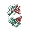
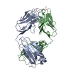
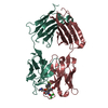
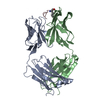
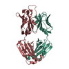
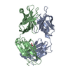
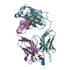
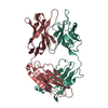
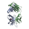
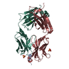
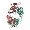
- PDB-4bh8: CRYSTAL STRUCTURE OF GERMLINE ANTIBODY 36-65 IN COMPLEX WITH PEPT... -
+ Open data

- Basic information
| Entry | Database: PDB / ID: 4bh8 | ||||||
|---|---|---|---|---|---|---|---|
| Title | CRYSTAL STRUCTURE OF GERMLINE ANTIBODY 36-65 IN COMPLEX WITH PEPTIDE GDPRPSYISHLL | ||||||
 Components Components |
| ||||||
 Keywords Keywords | IMMUNE SYSTEM / GERMLINE ANTIBODY / ANTIBODY POLYSPECIFICTY | ||||||
| Function / homology | Immunoglobulins / Immunoglobulin-like / Sandwich / Mainly Beta Function and homology information Function and homology information | ||||||
| Biological species |  SYNTHETIC CONSTRUCT (others) | ||||||
| Method |  X-RAY DIFFRACTION / MOLECULAR REPLACEMENT / Resolution: 2.4 Å X-RAY DIFFRACTION / MOLECULAR REPLACEMENT / Resolution: 2.4 Å | ||||||
 Authors Authors | Khan, T. / Salunke, D.M. | ||||||
 Citation Citation | Journal: J.Immunol. / Year: 2014 Title: Adjustable Locks and Flexible Keys: Plasticity of Epitope-Paratope Interactions in Germline Antibodies. Authors: Khan, T. / Salunke, D.M. | ||||||
| History |
|
- Structure visualization
| Structure viewer | Molecule: MolmilJmol/JSmol |
|---|
- Downloads & links
-Download
| PDBx/mmCIF format | 4bh8.cif.gz | 100.4 KB | Display | PDBx/mmCIF format |
|---|---|---|---|---|
| PDB format | pdb4bh8.ent.gz | 76.6 KB | Display | PDB format |
| PDBx/mmJSON format | 4bh8.json.gz | Tree view | PDBx/mmJSON format | |
| Others |  Other downloads Other downloads |
-Validation report
| Arichive directory | https://data.pdbj.org/pub/pdb/validation_reports/bh/4bh8ftp://data.pdbj.org/pub/pdb/validation_reports/bh/4bh8 | HTTPS FTP |
|---|
-Related structure data
| Related structure data | 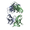 4bh7C 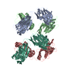 2a6jS C: citing same article ( S: Starting model for refinement |
|---|---|
| Similar structure data |


-Links
 PDBj
PDBj

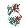
- Assembly
| Deposited unit | 
| ||||||||
|---|---|---|---|---|---|---|---|---|---|
| 1 |
| ||||||||
| Unit cell |
|
-Components
| #1: Antibody | Mass: 23663.084 Da / Num. of mol.: 1 / Fragment: ANTIGEN BINDING FRAGMENT / Source method: isolated from a natural source / Source: (natural) |
|---|---|
| #2: Antibody | Mass: 23942.768 Da / Num. of mol.: 1 / Fragment: ANTIGEN BINDING FRAGMENT / Source method: isolated from a natural source / Source: (natural) |
| #3: Protein/peptide | Mass: 1356.528 Da / Num. of mol.: 1 / Source method: obtained synthetically / Source: (synth.) SYNTHETIC CONSTRUCT (others) |
| #4: Water | ChemComp-HOH /  Mass: 18.015 Da / Num. of mol.: 190 / Source method: isolated from a natural source / Formula: H2O Mass: 18.015 Da / Num. of mol.: 190 / Source method: isolated from a natural source / Formula: H2O |
| Has protein modification | Y |
-Experimental details
-Experiment
| Experiment | Method: X-RAY DIFFRACTION / Number of used crystals: 1 |
|---|
- Sample preparation
| Crystal | Density Matthews: 2.43 Å3/Da / Density % sol: 49.4 % / Description: NO |
|---|---|
| Crystal grow | pH: 6.7 / Details: 0.05 M SODIUM CACODYLATE, PH 6.7, 21% PEG |
-Data collection
| Diffraction | Mean temperature: 110 K |
|---|---|
| Diffraction source | Source: ROTATING ANODE / Type: RIGAKU RU300 / Wavelength: 1.5418 |
| Detector | Type: MARRESEARCH / Detector: IMAGE PLATE / Date: Apr 9, 2009 / Details: MIRRORS |
| Radiation | Monochromator: NI FILTER CMF12 38CU-6 / Protocol: SINGLE WAVELENGTH / Monochromatic (M) / Laue (L): M / Scattering type: x-ray |
| Radiation wavelength | Wavelength: 1.5418 Å / Relative weight: 1 |
| Reflection | Resolution: 2.4→58 Å / Num. obs: 19076 / % possible obs: 100 % / Observed criterion σ(I): 0 / Redundancy: 4.5 % / Biso Wilson estimate: 32 Å2 / Rmerge(I) obs: 0.1 / Net I/σ(I): 12 |
| Reflection shell | Resolution: 2.4→2.5 Å / Redundancy: 4.5 % / Rmerge(I) obs: 0.4 / Mean I/σ(I) obs: 3.2 / % possible all: 100 |
- Processing
| Software |
| ||||||||||||||||||||||||||||||||||||||||||||||||||||||||||||
|---|---|---|---|---|---|---|---|---|---|---|---|---|---|---|---|---|---|---|---|---|---|---|---|---|---|---|---|---|---|---|---|---|---|---|---|---|---|---|---|---|---|---|---|---|---|---|---|---|---|---|---|---|---|---|---|---|---|---|---|---|---|
| Refinement | Method to determine structure: MOLECULAR REPLACEMENT Starting model: PDB ENTRY 2A6J Resolution: 2.4→50 Å / Rfactor Rfree error: 0.006 / Data cutoff high absF: 10000 / Data cutoff low absF: 0 / Cross valid method: THROUGHOUT / σ(F): 0 / Stereochemistry target values: MAXIMUM LIKELIHOOD
| ||||||||||||||||||||||||||||||||||||||||||||||||||||||||||||
| Solvent computation | Solvent model: FLAT MODEL / Bsol: 21.262 Å2 / ksol: 0.4 e/Å3 | ||||||||||||||||||||||||||||||||||||||||||||||||||||||||||||
| Displacement parameters | Biso mean: 31 Å2
| ||||||||||||||||||||||||||||||||||||||||||||||||||||||||||||
| Refine analyze |
| ||||||||||||||||||||||||||||||||||||||||||||||||||||||||||||
| Refinement step | Cycle: LAST / Resolution: 2.4→50 Å
| ||||||||||||||||||||||||||||||||||||||||||||||||||||||||||||
| Refine LS restraints |
| ||||||||||||||||||||||||||||||||||||||||||||||||||||||||||||
| LS refinement shell | Resolution: 2.4→2.55 Å / Rfactor Rfree error: 0.021 / Total num. of bins used: 6
| ||||||||||||||||||||||||||||||||||||||||||||||||||||||||||||
| Xplor file |
|
