[English] 日本語
 Yorodumi
- PDB-4bfc: Crystal structure of the C-terminal CMP-Kdo binding domain of Waa... -
+ Open data
- Basic information
| Entry | Database: PDB / ID: 4bfc | ||||||
|---|---|---|---|---|---|---|---|
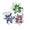
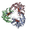
| Title | Crystal structure of the C-terminal CMP-Kdo binding domain of WaaA from Acinetobacter baumannii | ||||||
 Components Components | 3-DEOXY-D-MANNO-OCTULOSONIC-ACID TRANSFERASE | ||||||
 Keywords Keywords | TRANSFERASE | ||||||
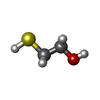
| Function / homology | Glycogen Phosphorylase B; / Rossmann fold / 3-Layer(aba) Sandwich / Alpha Beta / BETA-MERCAPTOETHANOL / 3-Deoxy-D-manno-octulosonic-acid transferase (Kdotransferase) family protein / :  Function and homology information Function and homology information | ||||||
| Biological species |  ACINETOBACTER BAUMANNII (bacteria) ACINETOBACTER BAUMANNII (bacteria) | ||||||
| Method |  X-RAY DIFFRACTION / X-RAY DIFFRACTION /  SYNCHROTRON / SYNCHROTRON /  MOLECULAR REPLACEMENT / Resolution: 1.7 Å MOLECULAR REPLACEMENT / Resolution: 1.7 Å | ||||||
 Authors Authors | Kimbung, Y.R. / Hakansson, M. / Logan, D. / Wang, P.F. / Schulz, M. / Mamat, U. / Woodard, R.W. | ||||||
 Citation Citation |  Journal: To be Published Journal: To be PublishedTitle: Crystal Structure of the C-Terminal Cmp-Kdo Binding Domain of Waaa from Acinetobacter Baumannii Authors: Kimbung, Y.R. / Hakansson, M. / Logan, D. / Wang, P.F. / Schulz, M. / Mamat, U. / Woodard, R.W. | ||||||
| History |
|
- Structure visualization
| Structure viewer | Molecule:  Molmil Molmil Jmol/JSmol Jmol/JSmol |
|---|
- Downloads & links
- Download
| PDBx/mmCIF format |  4bfc.cif.gz 4bfc.cif.gz | 59.7 KB | Display |  PDBx/mmCIF format PDBx/mmCIF format |
|---|---|---|---|---|
| PDB format |  pdb4bfc.ent.gz pdb4bfc.ent.gz | 41.6 KB | Display |  PDB format PDB format |
| PDBx/mmJSON format |  4bfc.json.gz 4bfc.json.gz | Tree view |  PDBx/mmJSON format PDBx/mmJSON format | |
| Others |  Other downloads Other downloads |
-Validation report
| Arichive directory |  https://data.pdbj.org/pub/pdb/validation_reports/bf/4bfc https://data.pdbj.org/pub/pdb/validation_reports/bf/4bfc ftp://data.pdbj.org/pub/pdb/validation_reports/bf/4bfc ftp://data.pdbj.org/pub/pdb/validation_reports/bf/4bfc | HTTPS FTP |
|---|
-Related structure data
| Related structure data |  2xciS S: Starting model for refinement |
|---|---|
| Similar structure data |
- Links
Links
- Assembly
| Deposited unit | 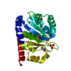
| ||||||||
|---|---|---|---|---|---|---|---|---|---|
| 1 | 
| ||||||||
| Unit cell |
|
- Components
| #1: Protein | Mass: 26774.201 Da / Num. of mol.: 1 Fragment: C-TERMINAL CMP-KDO BINDING DOMAIN, RESIDUES 220-430 Source method: isolated from a genetically manipulated source Source: (gene. exp.)  ACINETOBACTER BAUMANNII (bacteria) / Production host: ACINETOBACTER BAUMANNII (bacteria) / Production host:  | ||
|---|---|---|---|
| #2: Chemical | ChemComp-SO4 / | ||
| #3: Chemical | | #4: Water | ChemComp-HOH / | |
-Experimental details
-Experiment
| Experiment | Method:  X-RAY DIFFRACTION / Number of used crystals: 1 X-RAY DIFFRACTION / Number of used crystals: 1 |
|---|
- Sample preparation
| Crystal | Density Matthews: 3.46 Å3/Da / Density % sol: 64.5 % / Description: NONE |
|---|---|
| Crystal grow | pH: 6 Details: 0.1M NA CITRATE BUFFER PH 6.0, 0.2 MM LITHIUM SULPHATE AND 17 TO 23 % PEG 3350 |
-Data collection
| Diffraction | Mean temperature: 100 K | |||||||||||||||
|---|---|---|---|---|---|---|---|---|---|---|---|---|---|---|---|---|
| Diffraction source | Source:  SYNCHROTRON / Site: SYNCHROTRON / Site:  MAX II MAX II  / Beamline: I911-3 / Wavelength: 1.041 / Beamline: I911-3 / Wavelength: 1.041 | |||||||||||||||
| Detector | Type: MARRESEARCH MAR165 / Detector: CCD / Date: Feb 1, 2012 / Details: MIRROR | |||||||||||||||
| Radiation | Protocol: SINGLE WAVELENGTH / Monochromatic (M) / Laue (L): M / Scattering type: x-ray | |||||||||||||||
| Radiation wavelength | Wavelength: 1.041 Å / Relative weight: 1 | |||||||||||||||
| Reflection twin |
| |||||||||||||||
| Reflection | Resolution: 1.7→30 Å / Num. obs: 35057 / % possible obs: 99.4 % / Observed criterion σ(I): 3.77 / Redundancy: 5.7 % / Rmerge(I) obs: 0.05 / Net I/σ(I): 24.9 | |||||||||||||||
| Reflection shell | Resolution: 1.7→1.8 Å / Redundancy: 5.6 % / Rmerge(I) obs: 0.46 / Mean I/σ(I) obs: 3.77 / % possible all: 98.8 |
- Processing
| Software |
| ||||||||||||||||||||||||||||||||||||||||||||||||||||||||||||||||||||||||||||||||||||||||||||||||||||||||||||||||||||||||||||||||||||||||||||||||||||||||||||||||||||||||||||||||||||||
|---|---|---|---|---|---|---|---|---|---|---|---|---|---|---|---|---|---|---|---|---|---|---|---|---|---|---|---|---|---|---|---|---|---|---|---|---|---|---|---|---|---|---|---|---|---|---|---|---|---|---|---|---|---|---|---|---|---|---|---|---|---|---|---|---|---|---|---|---|---|---|---|---|---|---|---|---|---|---|---|---|---|---|---|---|---|---|---|---|---|---|---|---|---|---|---|---|---|---|---|---|---|---|---|---|---|---|---|---|---|---|---|---|---|---|---|---|---|---|---|---|---|---|---|---|---|---|---|---|---|---|---|---|---|---|---|---|---|---|---|---|---|---|---|---|---|---|---|---|---|---|---|---|---|---|---|---|---|---|---|---|---|---|---|---|---|---|---|---|---|---|---|---|---|---|---|---|---|---|---|---|---|---|---|
| Refinement | Method to determine structure:  MOLECULAR REPLACEMENT MOLECULAR REPLACEMENTStarting model: PDB ENTRY 2XCI Resolution: 1.7→28.33 Å / Cor.coef. Fo:Fc: 0.977 / Cor.coef. Fo:Fc free: 0.965 / SU B: 2.229 / SU ML: 0.062 / Cross valid method: THROUGHOUT / ESU R: 0.013 / ESU R Free: 0.014 / Stereochemistry target values: MAXIMUM LIKELIHOOD Details: HYDROGENS HAVE BEEN ADDED IN THE RIDING POSITIONS. U VALUES REFINED INDIVIDUALLY. HYDROGENS HAVE BEEN USED IF PRESENT IN THE INPUT SIDE CHAINS WITH POOR ELECTRON DENSITY WERE MODELED WITH ...Details: HYDROGENS HAVE BEEN ADDED IN THE RIDING POSITIONS. U VALUES REFINED INDIVIDUALLY. HYDROGENS HAVE BEEN USED IF PRESENT IN THE INPUT SIDE CHAINS WITH POOR ELECTRON DENSITY WERE MODELED WITH LOWER OCCUPANCY. MULTIPLE CONFORMERS WERE MODELED WITH LOWER OCCUPANCY ADDING UP TO 1.
| ||||||||||||||||||||||||||||||||||||||||||||||||||||||||||||||||||||||||||||||||||||||||||||||||||||||||||||||||||||||||||||||||||||||||||||||||||||||||||||||||||||||||||||||||||||||
| Solvent computation | Ion probe radii: 0.8 Å / Shrinkage radii: 0.8 Å / VDW probe radii: 1.2 Å / Solvent model: MASK | ||||||||||||||||||||||||||||||||||||||||||||||||||||||||||||||||||||||||||||||||||||||||||||||||||||||||||||||||||||||||||||||||||||||||||||||||||||||||||||||||||||||||||||||||||||||
| Displacement parameters | Biso mean: 29.23 Å2
| ||||||||||||||||||||||||||||||||||||||||||||||||||||||||||||||||||||||||||||||||||||||||||||||||||||||||||||||||||||||||||||||||||||||||||||||||||||||||||||||||||||||||||||||||||||||
| Refinement step | Cycle: LAST / Resolution: 1.7→28.33 Å
| ||||||||||||||||||||||||||||||||||||||||||||||||||||||||||||||||||||||||||||||||||||||||||||||||||||||||||||||||||||||||||||||||||||||||||||||||||||||||||||||||||||||||||||||||||||||
| Refine LS restraints |
|
 Movie
Movie Controller





 PDBj
PDBj


