 Movie
Movie Controller

[English] 日本語
 Yorodumi
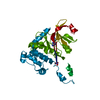
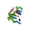
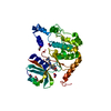
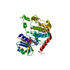
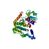
- PDB-4bdc: Fragment-based screening identifies a new area for inhibitor bind... -
+ Open data

- Basic information
| Entry | Database: PDB / ID: 4bdc | ||||||
|---|---|---|---|---|---|---|---|
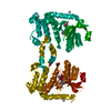
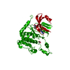
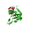
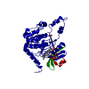
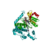
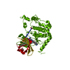
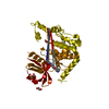
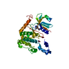
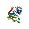
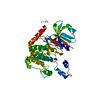
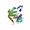
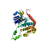
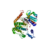
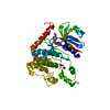
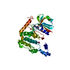
| Title | Fragment-based screening identifies a new area for inhibitor binding to checkpoint kinase 2 (CHK2) | ||||||
 Components Components | SERINE/THREONINE-PROTEIN KINASE CHK2 | ||||||
 Keywords Keywords | TRANSFERASE | ||||||
| Function / homology |  Function and homology information Function and homology informationpositive regulation of anoikis / mitotic DNA damage checkpoint signaling / cellular response to bisphenol A / regulation of autophagosome assembly / mitotic intra-S DNA damage checkpoint signaling / response to glycoside / thymocyte apoptotic process / cellular response to stress / negative regulation of DNA damage checkpoint / regulation of protein catabolic process ...positive regulation of anoikis / mitotic DNA damage checkpoint signaling / cellular response to bisphenol A / regulation of autophagosome assembly / mitotic intra-S DNA damage checkpoint signaling / response to glycoside / thymocyte apoptotic process / cellular response to stress / negative regulation of DNA damage checkpoint / regulation of protein catabolic process / replicative senescence / intrinsic apoptotic signaling pathway in response to DNA damage by p53 class mediator / mitotic spindle assembly / Chk1/Chk2(Cds1) mediated inactivation of Cyclin B:Cdk1 complex / signal transduction in response to DNA damage / DNA damage checkpoint signaling / regulation of signal transduction by p53 class mediator / protein catabolic process / DNA damage response, signal transduction by p53 class mediator / Stabilization of p53 / cellular response to gamma radiation / PML body / G2/M DNA damage checkpoint / Regulation of TP53 Activity through Methylation / Ubiquitin-Mediated Degradation of Phosphorylated Cdc25A / G2/M transition of mitotic cell cycle / cellular response to xenobiotic stimulus / intrinsic apoptotic signaling pathway in response to DNA damage / Regulation of TP53 Degradation / protein autophosphorylation / double-strand break repair / Recruitment and ATM-mediated phosphorylation of repair and signaling proteins at DNA double strand breaks / Regulation of TP53 Activity through Phosphorylation / protein phosphorylation / non-specific serine/threonine protein kinase / protein stabilization / protein serine kinase activity / cell division / protein serine/threonine kinase activity / DNA damage response / ubiquitin protein ligase binding / regulation of DNA-templated transcription / protein kinase binding / positive regulation of DNA-templated transcription / Golgi apparatus / protein homodimerization activity / nucleoplasm / ATP binding / metal ion binding / identical protein binding / nucleus / cytoplasm Similarity search - Function | ||||||
| Biological species |  HOMO SAPIENS (human) HOMO SAPIENS (human) | ||||||
| Method |  X-RAY DIFFRACTION / SYNCHROTRON / MOLECULAR REPLACEMENT / Resolution: 3 Å X-RAY DIFFRACTION / SYNCHROTRON / MOLECULAR REPLACEMENT / Resolution: 3 Å | ||||||
 Authors Authors | Silva-Santisteban, M.C. / Westwood, I.M. / Boxall, K. / Brown, N. / Peacock, S. / McAndrew, C. / Barrie, E. / Richards, M. / Mirza, A. / Oliver, A.W. ...Silva-Santisteban, M.C. / Westwood, I.M. / Boxall, K. / Brown, N. / Peacock, S. / McAndrew, C. / Barrie, E. / Richards, M. / Mirza, A. / Oliver, A.W. / Burke, R. / Hoelder, S. / Jones, K. / Aherne, G.W. / Blagg, J. / Collins, I. / Garrett, M.D. / van Montfort, R.L.M. | ||||||
 Citation Citation | Journal: Plos One / Year: 2013 Title: Fragment-Based Screening Maps Inhibitor Interactions in the ATP-Binding Site of Checkpoint Kinase 2. Authors: Silva-Santisteban, M.C. / Westwood, I.M. / Boxall, K. / Brown, N. / Peacock, S. / Mcandrew, C. / Barrie, E. / Richards, M. / Mirza, A. / Oliver, A.W. / Burke, R. / Hoelder, S. / Jones, K. / ...Authors: Silva-Santisteban, M.C. / Westwood, I.M. / Boxall, K. / Brown, N. / Peacock, S. / Mcandrew, C. / Barrie, E. / Richards, M. / Mirza, A. / Oliver, A.W. / Burke, R. / Hoelder, S. / Jones, K. / Aherne, G.W. / Blagg, J. / Collins, I. / Garrett, M.D. / Van Montfort, R.L. | ||||||
| History |
|
- Structure visualization
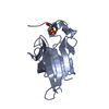
| Structure viewer | Molecule: MolmilJmol/JSmol |
|---|
- Downloads & links
-Download
| PDBx/mmCIF format | 4bdc.cif.gz | 126.5 KB | Display | PDBx/mmCIF format |
|---|---|---|---|---|
| PDB format | pdb4bdc.ent.gz | 97.5 KB | Display | PDB format |
| PDBx/mmJSON format | 4bdc.json.gz | Tree view | PDBx/mmJSON format | |
| Others |  Other downloads Other downloads |
-Validation report
| Arichive directory | https://data.pdbj.org/pub/pdb/validation_reports/bd/4bdcftp://data.pdbj.org/pub/pdb/validation_reports/bd/4bdc | HTTPS FTP |
|---|
-Related structure data
| Related structure data |  4bdaC 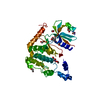 4bdbC  4bddC  4bdeC 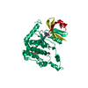 4bdfC  4bdgC 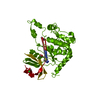 4bdhC 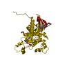 4bdiC 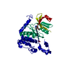 4bdjC  4bdkC  2wtjS S: Starting model for refinement C: citing same article ( |
|---|---|
| Similar structure data |


-Links
 PDBj
PDBj


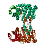
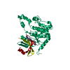
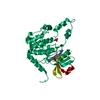
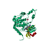
- Assembly
| Deposited unit | 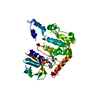
| ||||||||
|---|---|---|---|---|---|---|---|---|---|
| 1 |
| ||||||||
| Unit cell |
|
-Components
-Protein , 1 types, 1 molecules A
| #1: Protein | Mass: 37111.844 Da / Num. of mol.: 1 / Fragment: KINASE DOMAIN, RESIDUES 210-531 Source method: isolated from a genetically manipulated source Source: (gene. exp.) HOMO SAPIENS (human) / Plasmid: PTHREE-E / Production host:  References: UniProt: O96017, non-specific serine/threonine protein kinase |
|---|
-Non-polymers , 5 types, 19 molecules 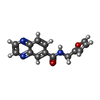


| #2: Chemical | ChemComp-ODH /  Mass: 253.256 Da / Num. of mol.: 1 / Source method: obtained synthetically / Formula: C14H11N3O2 Mass: 253.256 Da / Num. of mol.: 1 / Source method: obtained synthetically / Formula: C14H11N3O2 |
|---|---|
| #3: Chemical | ChemComp-NO3 /  Mass: 62.005 Da / Num. of mol.: 1 / Source method: obtained synthetically / Formula: NO3 Mass: 62.005 Da / Num. of mol.: 1 / Source method: obtained synthetically / Formula: NO3 |
| #4: Chemical | ChemComp-CL /  Mass: 35.453 Da / Num. of mol.: 1 / Source method: obtained synthetically / Formula: Cl Mass: 35.453 Da / Num. of mol.: 1 / Source method: obtained synthetically / Formula: Cl |
| #5: Chemical | ChemComp-EDO /  Mass: 62.068 Da / Num. of mol.: 1 / Source method: obtained synthetically / Formula: C2H6O2 Mass: 62.068 Da / Num. of mol.: 1 / Source method: obtained synthetically / Formula: C2H6O2 |
| #6: Water | ChemComp-HOH / Mass: 18.015 Da / Num. of mol.: 15 / Source method: isolated from a natural source / Formula: H2O |
-Experimental details
-Experiment
| Experiment | Method: X-RAY DIFFRACTION / Number of used crystals: 1 |
|---|
- Sample preparation
| Crystal | Density Matthews: 3.01 Å3/Da / Density % sol: 59.13 % / Description: NONE |
|---|---|
| Crystal grow | Details: 0.1 M HEPES 7.5, 0.2 M MG(NO3)2, 10% (V/V) ETHYLENE GLYCOL, 1 MM TCEP AND 8-14% (W/V) PEG 3350 |
-Data collection
| Diffraction | Mean temperature: 100 K |
|---|---|
| Diffraction source | Source: SYNCHROTRON / Site: Diamond  / Beamline: I04 / Wavelength: 0.9728 / Beamline: I04 / Wavelength: 0.9728 |
| Detector | Type: ADSC CCD / Detector: CCD / Date: Dec 17, 2008 |
| Radiation | Protocol: SINGLE WAVELENGTH / Monochromatic (M) / Laue (L): M / Scattering type: x-ray |
| Radiation wavelength | Wavelength: 0.9728 Å / Relative weight: 1 |
| Reflection | Resolution: 3→40.92 Å / Num. obs: 9297 / % possible obs: 99.9 % / Observed criterion σ(I): 1.5 / Redundancy: 6 % / Biso Wilson estimate: 95.93 Å2 / Rmerge(I) obs: 0.09 / Net I/σ(I): 11.2 |
| Reflection shell | Resolution: 3→3.16 Å / Redundancy: 6.2 % / Rmerge(I) obs: 0.49 / Mean I/σ(I) obs: 2 / % possible all: 100 |
- Processing
| Software |
| ||||||||||||||||||||||||||||||||||||||||||||||||||||||||||||||||||||||||||||||||||||||||||||||||||||||||||||||||||
|---|---|---|---|---|---|---|---|---|---|---|---|---|---|---|---|---|---|---|---|---|---|---|---|---|---|---|---|---|---|---|---|---|---|---|---|---|---|---|---|---|---|---|---|---|---|---|---|---|---|---|---|---|---|---|---|---|---|---|---|---|---|---|---|---|---|---|---|---|---|---|---|---|---|---|---|---|---|---|---|---|---|---|---|---|---|---|---|---|---|---|---|---|---|---|---|---|---|---|---|---|---|---|---|---|---|---|---|---|---|---|---|---|---|---|---|
| Refinement | Method to determine structure: MOLECULAR REPLACEMENT Starting model: PDB ENTRY 2WTJ Resolution: 3→40.92 Å / Cor.coef. Fo:Fc: 0.9504 / Cor.coef. Fo:Fc free: 0.9371 / SU R Cruickshank DPI: 1.053 / Cross valid method: THROUGHOUT / σ(F): 0 / SU R Blow DPI: 1.112 / SU Rfree Blow DPI: 0.306 / SU Rfree Cruickshank DPI: 0.31
| ||||||||||||||||||||||||||||||||||||||||||||||||||||||||||||||||||||||||||||||||||||||||||||||||||||||||||||||||||
| Displacement parameters | Biso mean: 78.82 Å2
| ||||||||||||||||||||||||||||||||||||||||||||||||||||||||||||||||||||||||||||||||||||||||||||||||||||||||||||||||||
| Refine analyze | Luzzati coordinate error obs: 0.38 Å | ||||||||||||||||||||||||||||||||||||||||||||||||||||||||||||||||||||||||||||||||||||||||||||||||||||||||||||||||||
| Refinement step | Cycle: LAST / Resolution: 3→40.92 Å
| ||||||||||||||||||||||||||||||||||||||||||||||||||||||||||||||||||||||||||||||||||||||||||||||||||||||||||||||||||
| Refine LS restraints |
| ||||||||||||||||||||||||||||||||||||||||||||||||||||||||||||||||||||||||||||||||||||||||||||||||||||||||||||||||||
| LS refinement shell | Resolution: 3→3.35 Å / Total num. of bins used: 5
| ||||||||||||||||||||||||||||||||||||||||||||||||||||||||||||||||||||||||||||||||||||||||||||||||||||||||||||||||||
| Refinement TLS params. | Method: refined / Origin x: -25.1366 Å / Origin y: 29.4605 Å / Origin z: 10.3202 Å
| ||||||||||||||||||||||||||||||||||||||||||||||||||||||||||||||||||||||||||||||||||||||||||||||||||||||||||||||||||
| Refinement TLS group | Selection details: CHAIN A |
