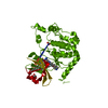
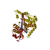
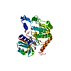
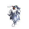
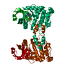
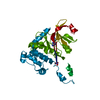
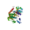
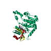
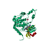
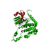
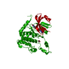
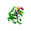
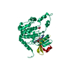
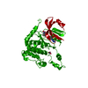
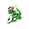
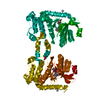
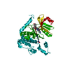
- PDB-4a9s: CRYSTAL STRUCTURE OF HUMAN CHK2 IN COMPLEX WITH BENZIMIDAZOLE CAR... -
+
Open data
ID or keywords:
Loading...
-
Basic information
Entry
Database: PDB / ID: 4a9s
Title
CRYSTAL STRUCTURE OF HUMAN CHK2 IN COMPLEX WITH BENZIMIDAZOLE CARBOXAMIDE INHIBITOR
Components
SERINE/THREONINE-PROTEIN KINASE CHK2
Keywords
TRANSFERASE
Function / homology
Function and homology information
positive regulation of anoikis / cellular response to bisphenol A / mitotic DNA damage checkpoint signaling / regulation of autophagosome assembly / mitotic intra-S DNA damage checkpoint signaling / response to glycoside / thymocyte apoptotic process / cellular response to stress / negative regulation of DNA damage checkpoint / regulation of protein catabolic process ...positive regulation of anoikis / cellular response to bisphenol A / mitotic DNA damage checkpoint signaling / regulation of autophagosome assembly / mitotic intra-S DNA damage checkpoint signaling / response to glycoside / thymocyte apoptotic process / cellular response to stress / negative regulation of DNA damage checkpoint / regulation of protein catabolic process / replicative senescence / mitotic spindle assembly / intrinsic apoptotic signaling pathway in response to DNA damage by p53 class mediator / Chk1/Chk2(Cds1) mediated inactivation of Cyclin B:Cdk1 complex / signal transduction in response to DNA damage / DNA damage checkpoint signaling / regulation of signal transduction by p53 class mediator / protein catabolic process / DNA damage response, signal transduction by p53 class mediator / Stabilization of p53 / cellular response to gamma radiation / cellular response to xenobiotic stimulus / PML body / G2/M DNA damage checkpoint / Regulation of TP53 Activity through Methylation / Ubiquitin-Mediated Degradation of Phosphorylated Cdc25A / intrinsic apoptotic signaling pathway in response to DNA damage / G2/M transition of mitotic cell cycle / Regulation of TP53 Degradation / protein autophosphorylation / double-strand break repair / Recruitment and ATM-mediated phosphorylation of repair and signaling proteins at DNA double strand breaks / Regulation of TP53 Activity through Phosphorylation / protein phosphorylation / non-specific serine/threonine protein kinase / protein stabilization / protein serine kinase activity / cell division / protein serine/threonine kinase activity / ubiquitin protein ligase binding / regulation of DNA-templated transcription / DNA damage response / protein kinase binding / positive regulation of DNA-templated transcription / Golgi apparatus / protein homodimerization activity / nucleoplasm / ATP binding / metal ion binding / identical protein binding / nucleus / cytoplasm Similarity search - Function
In the structure databanks used in Yorodumi, some data are registered as the other names, "COVID-19 virus" and "2019-nCoV". Here are the details of the virus and the list of structure data.
Jan 31, 2019. EMDB accession codes are about to change! (news from PDBe EMDB page)
EMDB accession codes are about to change! (news from PDBe EMDB page)
The allocation of 4 digits for EMDB accession codes will soon come to an end. Whilst these codes will remain in use, new EMDB accession codes will include an additional digit and will expand incrementally as the available range of codes is exhausted. The current 4-digit format prefixed with “EMD-” (i.e. EMD-XXXX) will advance to a 5-digit format (i.e. EMD-XXXXX), and so on. It is currently estimated that the 4-digit codes will be depleted around Spring 2019, at which point the 5-digit format will come into force.
The EM Navigator/Yorodumi systems omit the EMD- prefix.
Related info.:Q: What is EMD? / ID/Accession-code notation in Yorodumi/EM Navigator
Yorodumi is a browser for structure data from EMDB, PDB, SASBDB, etc.
This page is also the successor to EM Navigator detail page, and also detail information page/front-end page for Omokage search.
The word "yorodu" (or yorozu) is an old Japanese word meaning "ten thousand". "mi" (miru) is to see.
Related info.:EMDB / PDB / SASBDB / Comparison of 3 databanks / Yorodumi Search / Aug 31, 2016. New EM Navigator & Yorodumi / Yorodumi Papers / Jmol/JSmol / Function and homology information / Changes in new EM Navigator and Yorodumi
 Movie
Movie Controller

 Yorodumi
 Open data
 Basic information
 Components
 Keywords
 Function and homology information
 HOMO SAPIENS (human)
 X-RAY DIFFRACTION /
  Authors
 Citation
 Structure visualization
 Downloads & links
 Other downloads
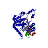


 PDBj
PDBj


 Assembly
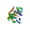


 Mass: 62.005 Da / Num. of mol.: 1 / Source method: obtained synthetically / Formula: NO3
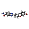
 Mass: 345.351 Da / Num. of mol.: 1 / Source method: obtained synthetically / Formula: C20H15N3O3
 Mass: 18.015 Da / Num. of mol.: 28 / Source method: isolated from a natural source / Formula: H2O
 Sample preparation
 / Beamline: I03 / Wavelength: 0.9763
  Processing