 Movie
Movie Controller

[English] 日本語
 Yorodumi
- PDB-4b9j: Structure of self-complemented CssA subunit of enterotoxigenic Es... -
+ Open data

- Basic information
| Entry | Database: PDB / ID: 4b9j | ||||||
|---|---|---|---|---|---|---|---|
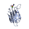
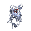
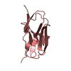
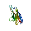
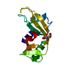
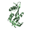
| Title | Structure of self-complemented CssA subunit of enterotoxigenic Escherichia coli colonization factor CS6 | ||||||
 Components Components | CS6 FIMBRIAL SUBUNIT A | ||||||
 Keywords Keywords | CELL ADHESION / DIARRHEAL DISEASE / FIMBRIAE / FUSION PROTEIN | ||||||
| Function / homology | Immunoglobulin-like - #3480 / : / pilus / Immunoglobulin-like / Sandwich / Mainly Beta / CS6 fimbrial subunit A Function and homology information Function and homology information | ||||||
| Biological species |  | ||||||
| Method |  X-RAY DIFFRACTION / SYNCHROTRON / MOLECULAR REPLACEMENT / Resolution: 2.542 Å X-RAY DIFFRACTION / SYNCHROTRON / MOLECULAR REPLACEMENT / Resolution: 2.542 Å | ||||||
 Authors Authors | Roy, S.P. / Rahman, M.M. / Yu, X.D. / Tuittila, M. / Knight, S.D. / Zavialov, A.V. | ||||||
 Citation Citation | Journal: Mol.Microbiol. / Year: 2012 Title: Crystal Structure of Enterotoxigenic Escherichia Coli Colonization Factor Cs6 Reveals a Novel Type of Functional Assembly. Authors: Roy, S.P. / Rahman, M.M. / Yu, X.D. / Tuittila, M. / Knight, S.D. / Zavialov, A.V. | ||||||
| History |
|
- Structure visualization
| Structure viewer | Molecule: MolmilJmol/JSmol |
|---|
- Downloads & links
-Download
| PDBx/mmCIF format | 4b9j.cif.gz | 36.3 KB | Display | PDBx/mmCIF format |
|---|---|---|---|---|
| PDB format | pdb4b9j.ent.gz | 24.6 KB | Display | PDB format |
| PDBx/mmJSON format | 4b9j.json.gz | Tree view | PDBx/mmJSON format | |
| Others |  Other downloads Other downloads |
-Validation report
| Arichive directory | https://data.pdbj.org/pub/pdb/validation_reports/b9/4b9jftp://data.pdbj.org/pub/pdb/validation_reports/b9/4b9j | HTTPS FTP |
|---|
-Related structure data
| Related structure data |  4b9gC 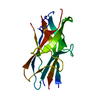 4b9iSC C: citing same article ( S: Starting model for refinement |
|---|---|
| Similar structure data |


-Links
 PDBj
PDBj- Assembly
| Deposited unit | 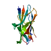
| |||||||||
|---|---|---|---|---|---|---|---|---|---|---|
| 1 |
| |||||||||
| Unit cell |
| |||||||||
| Components on special symmetry positions |
|
-Components
| #1: Protein | Mass: 16643.484 Da / Num. of mol.: 1 Source method: isolated from a genetically manipulated source Details: LINKER SEQUENCE DNKQ / Source: (gene. exp.) |
|---|---|
| #2: Water | ChemComp-HOH /  Mass: 18.015 Da / Num. of mol.: 28 / Source method: isolated from a natural source / Formula: H2O Mass: 18.015 Da / Num. of mol.: 28 / Source method: isolated from a natural source / Formula: H2O |
-Experimental details
-Experiment
| Experiment | Method: X-RAY DIFFRACTION / Number of used crystals: 1 |
|---|
- Sample preparation
| Crystal | Density Matthews: 1.78 Å3/Da / Density % sol: 31 % / Description: NONE |
|---|---|
| Crystal grow | pH: 4.6 Details: 24-30 % PEG4000 IN 0.2 M AMMONIUM ACETATE, 0.1 M NA ACETATE, PH 4.6 |
-Data collection
| Diffraction | Mean temperature: 287 K |
|---|---|
| Diffraction source | Source: SYNCHROTRON / Site: ESRF  / Beamline: BM14 / Wavelength: 0.9334 / Beamline: BM14 / Wavelength: 0.9334 |
| Detector | Type: MARRESEARCH / Detector: CCD / Date: Feb 24, 2011 / Details: BAND PASS 1.9X10⁻⁴ FOR A SI(111) MONOCHROMATOR |
| Radiation | Monochromator: SI(111) MONOCHROMATOR / Protocol: SINGLE WAVELENGTH / Monochromatic (M) / Laue (L): M / Scattering type: x-ray |
| Radiation wavelength | Wavelength: 0.9334 Å / Relative weight: 1 |
| Reflection | Resolution: 2.54→43 Å / Num. obs: 4190 / % possible obs: 98.8 % / Observed criterion σ(I): 2 / Redundancy: 3.2 % / Biso Wilson estimate: 37.719 Å2 / Rmerge(I) obs: 0.03 / Net I/σ(I): 33.9 |
| Reflection shell | Resolution: 2.54→2.68 Å / Redundancy: 3.2 % / Rmerge(I) obs: 0.07 / Mean I/σ(I) obs: 16.8 / % possible all: 98.3 |
- Processing
| Software |
| ||||||||||||||||||||||||
|---|---|---|---|---|---|---|---|---|---|---|---|---|---|---|---|---|---|---|---|---|---|---|---|---|---|
| Refinement | Method to determine structure: MOLECULAR REPLACEMENT Starting model: PDB ENTRY 4B9I Resolution: 2.542→37.125 Å / SU ML: 0.27 / σ(F): 0 / Phase error: 27.1 / Stereochemistry target values: ML
| ||||||||||||||||||||||||
| Solvent computation | Shrinkage radii: 0.95 Å / VDW probe radii: 1.2 Å / Solvent model: FLAT BULK SOLVENT MODEL / Bsol: 32.603 Å2 / ksol: 0.355 e/Å3 | ||||||||||||||||||||||||
| Displacement parameters |
| ||||||||||||||||||||||||
| Refinement step | Cycle: LAST / Resolution: 2.542→37.125 Å
| ||||||||||||||||||||||||
| Refine LS restraints |
| ||||||||||||||||||||||||
| LS refinement shell | Resolution: 2.5423→37.1289 Å
|
