 Movie
Movie Controller


+ Open data

- Basic information
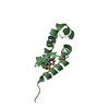
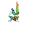
| Entry | Database: PDB / ID: 4b6d | ||||||
|---|---|---|---|---|---|---|---|
| Title | Structure of the atypical C1 domain of MgcRacGAP | ||||||
 Components Components | RAC GTPASE-ACTIVATING PROTEIN 1 | ||||||
 Keywords Keywords | SIGNALING PROTEIN / CYTOKINESIS / PLASMA MEMBRANE / PHOSPHOLIPIDS / CENTRALSPINDLIN / SPINDLE MIDZONE / CENTRAL SPINDLE / MIDBODY | ||||||
| Function / homology |  Function and homology information Function and homology informationcentralspindlin complex / actomyosin contractile ring assembly / mitotic spindle midzone assembly / sulfate transmembrane transport / regulation of attachment of spindle microtubules to kinetochore / Flemming body / gamma-tubulin binding / RHOD GTPase cycle / Kinesins / regulation of small GTPase mediated signal transduction ...centralspindlin complex / actomyosin contractile ring assembly / mitotic spindle midzone assembly / sulfate transmembrane transport / regulation of attachment of spindle microtubules to kinetochore / Flemming body / gamma-tubulin binding / RHOD GTPase cycle / Kinesins / regulation of small GTPase mediated signal transduction / COPI-dependent Golgi-to-ER retrograde traffic / RHOB GTPase cycle / spindle midzone / positive regulation of cytokinesis / RHOC GTPase cycle / cleavage furrow / phosphatidylinositol-3,4,5-trisphosphate binding / mitotic cytokinesis / CDC42 GTPase cycle / neuroblast proliferation / regulation of embryonic development / RAC3 GTPase cycle / RHOA GTPase cycle / RAC2 GTPase cycle / alpha-tubulin binding / beta-tubulin binding / Rho protein signal transduction / RAC1 GTPase cycle / monoatomic ion transport / MHC class II antigen presentation / acrosomal vesicle / GTPase activator activity / erythrocyte differentiation / spindle / cytoplasmic side of plasma membrane / mitotic spindle / midbody / spermatogenesis / microtubule binding / microtubule / protein-macromolecule adaptor activity / protein kinase binding / mitochondrion / extracellular exosome / zinc ion binding / nucleoplasm / nucleus / cytosol Similarity search - Function | ||||||
| Biological species |  HOMO SAPIENS (human) HOMO SAPIENS (human) | ||||||
| Method |  X-RAY DIFFRACTION / SYNCHROTRON / SAD / Resolution: 2.2 Å X-RAY DIFFRACTION / SYNCHROTRON / SAD / Resolution: 2.2 Å | ||||||
 Authors Authors | Pye, V.E. / Lekomtsev, S. / Petronczki, M. / Cherepanov, P. | ||||||
 Citation Citation | Journal: Nature / Year: 2012 Title: Centralspindlin Links the Mitotic Spindle to the Plasma Membrane During Cytokinesis. Authors: Lekomtsev, S. / Su, K. / Pye, V.E. / Blight, K. / Sundaramoorthy, S. / Takaki, T. / Collinson, L.M. / Cherepanov, P. / Divecha, N. / Petronczki, M. | ||||||
| History |
|
- Structure visualization
| Structure viewer | Molecule: MolmilJmol/JSmol |
|---|
- Downloads & links
-Download
| PDBx/mmCIF format | 4b6d.cif.gz | 156.6 KB | Display | PDBx/mmCIF format |
|---|---|---|---|---|
| PDB format | pdb4b6d.ent.gz | 125.6 KB | Display | PDB format |
| PDBx/mmJSON format | 4b6d.json.gz | Tree view | PDBx/mmJSON format | |
| Others |  Other downloads Other downloads |
-Validation report
| Arichive directory | https://data.pdbj.org/pub/pdb/validation_reports/b6/4b6dftp://data.pdbj.org/pub/pdb/validation_reports/b6/4b6d | HTTPS FTP |
|---|
-Related structure data
| Related structure data |  1ptqS S: Starting model for refinement |
|---|---|
| Similar structure data |


-Links
 PDBj
PDBj


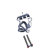
- Assembly
| Deposited unit | 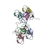
| ||||||||||||||||||||||||
|---|---|---|---|---|---|---|---|---|---|---|---|---|---|---|---|---|---|---|---|---|---|---|---|---|---|
| 1 | 
| ||||||||||||||||||||||||
| 2 | 
| ||||||||||||||||||||||||
| 3 | 
| ||||||||||||||||||||||||
| 4 | 
| ||||||||||||||||||||||||
| 5 | 
| ||||||||||||||||||||||||
| 6 | 
| ||||||||||||||||||||||||
| Unit cell |
| ||||||||||||||||||||||||
| Noncrystallographic symmetry (NCS) | NCS oper:
|
-Components
| #1: Protein | Mass: 6823.265 Da / Num. of mol.: 6 / Fragment: C1 DOMAIN, RESIDUES 284-339 Source method: isolated from a genetically manipulated source Source: (gene. exp.) HOMO SAPIENS (human) / Plasmid: PGEX-C1WT / Production host:  #2: Chemical | ChemComp-GOL / |   Mass: 92.094 Da / Num. of mol.: 1 / Source method: obtained synthetically / Formula: C3H8O3 Mass: 92.094 Da / Num. of mol.: 1 / Source method: obtained synthetically / Formula: C3H8O3#3: Chemical | ChemComp-ZN /   Mass: 65.409 Da / Num. of mol.: 12 / Source method: obtained synthetically / Formula: Zn Mass: 65.409 Da / Num. of mol.: 12 / Source method: obtained synthetically / Formula: Zn#4: Water | ChemComp-HOH / |  Mass: 18.015 Da / Num. of mol.: 163 / Source method: isolated from a natural source / Formula: H2O Mass: 18.015 Da / Num. of mol.: 163 / Source method: isolated from a natural source / Formula: H2OSequence details | RESIDUES 284-338 WITH AN N-TERMINAL EXTENSION SEQUENCE EXTENSION SEQUENCE GPLGS | |
|---|
-Experimental details
-Experiment
| Experiment | Method: X-RAY DIFFRACTION / Number of used crystals: 1 |
|---|
- Sample preparation
| Crystal | Density Matthews: 3.35 Å3/Da / Density % sol: 63.26 % / Description: NONE |
|---|---|
| Crystal grow | pH: 7.1 Details: 1.3 M SODIUM CITRATE, PH 7.1 AND 0.3 M DIMETHYLETHYLAMMONIUM PROPANE SULFONATE |
-Data collection
| Diffraction | Mean temperature: 100 K |
|---|---|
| Diffraction source | Source: SYNCHROTRON / Site: Diamond  / Beamline: I02 / Wavelength: 0.9795 / Beamline: I02 / Wavelength: 0.9795 |
| Detector | Type: DECTRIS PILATUS 6M / Detector: PIXEL / Date: Jul 31, 2012 |
| Radiation | Protocol: SINGLE WAVELENGTH / Monochromatic (M) / Laue (L): M / Scattering type: x-ray |
| Radiation wavelength | Wavelength: 0.9795 Å / Relative weight: 1 |
| Reflection twin | Operator: H,-K,-H-L / Fraction: 0.5 |
| Reflection | Resolution: 2.2→66.93 Å / Num. obs: 26140 / % possible obs: 99.4 % / Observed criterion σ(I): 2.6 / Redundancy: 9.7 % / Biso Wilson estimate: 33.3 Å2 / Rmerge(I) obs: 0.15 / Net I/σ(I): 9.4 |
| Reflection shell | Resolution: 2.2→2.32 Å / Redundancy: 8.9 % / Rmerge(I) obs: 0.96 / Mean I/σ(I) obs: 2.6 / % possible all: 98.8 |
- Processing
| Software |
| |||||||||||||||||||||||||||||||||||||||||||||||||||||||||||||||||||||||||||||||||||||||||||||||||||||||||||||||||||||||||||||||||||||||||||||||||||||||||||||||||||||||||||||||||||||||||||||||||||||||||||||||||||||||||||||||||||||||||||||||||||||||||||||||||||||||||||||||||||||||||||||||||||||||||||||||||||||||||||||||||||||
|---|---|---|---|---|---|---|---|---|---|---|---|---|---|---|---|---|---|---|---|---|---|---|---|---|---|---|---|---|---|---|---|---|---|---|---|---|---|---|---|---|---|---|---|---|---|---|---|---|---|---|---|---|---|---|---|---|---|---|---|---|---|---|---|---|---|---|---|---|---|---|---|---|---|---|---|---|---|---|---|---|---|---|---|---|---|---|---|---|---|---|---|---|---|---|---|---|---|---|---|---|---|---|---|---|---|---|---|---|---|---|---|---|---|---|---|---|---|---|---|---|---|---|---|---|---|---|---|---|---|---|---|---|---|---|---|---|---|---|---|---|---|---|---|---|---|---|---|---|---|---|---|---|---|---|---|---|---|---|---|---|---|---|---|---|---|---|---|---|---|---|---|---|---|---|---|---|---|---|---|---|---|---|---|---|---|---|---|---|---|---|---|---|---|---|---|---|---|---|---|---|---|---|---|---|---|---|---|---|---|---|---|---|---|---|---|---|---|---|---|---|---|---|---|---|---|---|---|---|---|---|---|---|---|---|---|---|---|---|---|---|---|---|---|---|---|---|---|---|---|---|---|---|---|---|---|---|---|---|---|---|---|---|---|---|---|---|---|---|---|---|---|---|---|---|---|---|---|---|---|---|---|---|---|---|---|---|---|---|---|---|---|---|---|---|---|---|---|---|---|---|---|---|---|---|---|---|---|---|---|---|---|---|---|---|---|---|---|---|---|---|---|---|---|---|---|---|
| Refinement | Method to determine structure: SAD Starting model: PDB ENTRY 1PTQ Resolution: 2.2→66.925 Å / σ(F): 1.14 / Phase error: 28.33 / Stereochemistry target values: TWIN_LSQ_F / Details: CHAINS A, B, C, D, E AND F ARE RELATED BY NCS.
| |||||||||||||||||||||||||||||||||||||||||||||||||||||||||||||||||||||||||||||||||||||||||||||||||||||||||||||||||||||||||||||||||||||||||||||||||||||||||||||||||||||||||||||||||||||||||||||||||||||||||||||||||||||||||||||||||||||||||||||||||||||||||||||||||||||||||||||||||||||||||||||||||||||||||||||||||||||||||||||||||||||
| Solvent computation | Shrinkage radii: 0.9 Å / VDW probe radii: 1.11 Å / Solvent model: FLAT BULK SOLVENT MODEL / Bsol: 0 Å2 / ksol: 0 e/Å3 | |||||||||||||||||||||||||||||||||||||||||||||||||||||||||||||||||||||||||||||||||||||||||||||||||||||||||||||||||||||||||||||||||||||||||||||||||||||||||||||||||||||||||||||||||||||||||||||||||||||||||||||||||||||||||||||||||||||||||||||||||||||||||||||||||||||||||||||||||||||||||||||||||||||||||||||||||||||||||||||||||||||
| Displacement parameters | Biso mean: 56.8 Å2 | |||||||||||||||||||||||||||||||||||||||||||||||||||||||||||||||||||||||||||||||||||||||||||||||||||||||||||||||||||||||||||||||||||||||||||||||||||||||||||||||||||||||||||||||||||||||||||||||||||||||||||||||||||||||||||||||||||||||||||||||||||||||||||||||||||||||||||||||||||||||||||||||||||||||||||||||||||||||||||||||||||||
| Refinement step | Cycle: LAST / Resolution: 2.2→66.925 Å
| |||||||||||||||||||||||||||||||||||||||||||||||||||||||||||||||||||||||||||||||||||||||||||||||||||||||||||||||||||||||||||||||||||||||||||||||||||||||||||||||||||||||||||||||||||||||||||||||||||||||||||||||||||||||||||||||||||||||||||||||||||||||||||||||||||||||||||||||||||||||||||||||||||||||||||||||||||||||||||||||||||||
| Refine LS restraints |
| |||||||||||||||||||||||||||||||||||||||||||||||||||||||||||||||||||||||||||||||||||||||||||||||||||||||||||||||||||||||||||||||||||||||||||||||||||||||||||||||||||||||||||||||||||||||||||||||||||||||||||||||||||||||||||||||||||||||||||||||||||||||||||||||||||||||||||||||||||||||||||||||||||||||||||||||||||||||||||||||||||||
| LS refinement shell |
| |||||||||||||||||||||||||||||||||||||||||||||||||||||||||||||||||||||||||||||||||||||||||||||||||||||||||||||||||||||||||||||||||||||||||||||||||||||||||||||||||||||||||||||||||||||||||||||||||||||||||||||||||||||||||||||||||||||||||||||||||||||||||||||||||||||||||||||||||||||||||||||||||||||||||||||||||||||||||||||||||||||
| Refinement TLS params. | Method: refined / Refine-ID: X-RAY DIFFRACTION
| |||||||||||||||||||||||||||||||||||||||||||||||||||||||||||||||||||||||||||||||||||||||||||||||||||||||||||||||||||||||||||||||||||||||||||||||||||||||||||||||||||||||||||||||||||||||||||||||||||||||||||||||||||||||||||||||||||||||||||||||||||||||||||||||||||||||||||||||||||||||||||||||||||||||||||||||||||||||||||||||||||||
| Refinement TLS group |
|
