 Movie
Movie Controller


+ Open data

- Basic information
| Entry | Database: PDB / ID: 3mp9 | ||||||
|---|---|---|---|---|---|---|---|
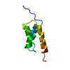
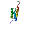
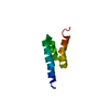
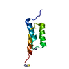
| Title | Structure of Streptococcal protein G B1 domain at pH 3.0 | ||||||
 Components Components | Immunoglobulin G-binding protein G | ||||||
 Keywords Keywords | PROTEIN BINDING / PROTEIN G / IGG-BINDING PROTEIN | ||||||
| Function / homology |  Function and homology information Function and homology information | ||||||
| Biological species |  Streptococcus sp. 'group G' (bacteria) Streptococcus sp. 'group G' (bacteria) | ||||||
| Method |  X-RAY DIFFRACTION / SYNCHROTRON / MOLECULAR REPLACEMENT / Resolution: 1.2 Å X-RAY DIFFRACTION / SYNCHROTRON / MOLECULAR REPLACEMENT / Resolution: 1.2 Å | ||||||
 Authors Authors | Tomlinson, J.H. / Green, V.L. / Baker, P.J. / Williamson, M.P. | ||||||
 Citation Citation | Journal: Proteins / Year: 2010 Title: Structural origins of pH-dependent chemical shifts in the B1 domain of protein G. Authors: Tomlinson, J.H. / Green, V.L. / Baker, P.J. / Williamson, M.P. | ||||||
| History |
|
- Structure visualization
| Structure viewer | Molecule: MolmilJmol/JSmol |
|---|
- Downloads & links
-Download
| PDBx/mmCIF format | 3mp9.cif.gz | 64.9 KB | Display | PDBx/mmCIF format |
|---|---|---|---|---|
| PDB format | pdb3mp9.ent.gz | 48.5 KB | Display | PDB format |
| PDBx/mmJSON format | 3mp9.json.gz | Tree view | PDBx/mmJSON format | |
| Others |  Other downloads Other downloads |
-Validation report
| Arichive directory | https://data.pdbj.org/pub/pdb/validation_reports/mp/3mp9ftp://data.pdbj.org/pub/pdb/validation_reports/mp/3mp9 | HTTPS FTP |
|---|
-Related structure data
| Related structure data | 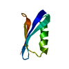 1pgaS S: Starting model for refinement |
|---|---|
| Similar structure data |


-Links
 PDBj
PDBj

- Assembly
| Deposited unit | 
| ||||||||
|---|---|---|---|---|---|---|---|---|---|
| 1 | 
| ||||||||
| 2 | 
| ||||||||
| 3 | 
| ||||||||
| Unit cell |
|
-Components
| #1: Antibody | Mass: 7216.829 Da / Num. of mol.: 2 / Fragment: B1 domain (UNP residues 227-282) Source method: isolated from a genetically manipulated source Source: (gene. exp.) Streptococcus sp. 'group G' (bacteria) / Gene: spg / Plasmid: pET15b / Production host: #2: Chemical |   Mass: 46.025 Da / Num. of mol.: 2 / Source method: obtained synthetically / Formula: CH2O2 Mass: 46.025 Da / Num. of mol.: 2 / Source method: obtained synthetically / Formula: CH2O2#3: Water | ChemComp-HOH / |  Mass: 18.015 Da / Num. of mol.: 87 / Source method: isolated from a natural source / Formula: H2O Mass: 18.015 Da / Num. of mol.: 87 / Source method: isolated from a natural source / Formula: H2O |
|---|
-Experimental details
-Experiment
| Experiment | Method: X-RAY DIFFRACTION / Number of used crystals: 1 |
|---|
- Sample preparation
| Crystal | Density Matthews: 1.74 Å3/Da / Density % sol: 29.3 % |
|---|---|
| Crystal grow | Temperature: 290 K / Method: vapor diffusion, hanging drop / pH: 3 Details: Crystals grown in 3.8M sodium formate, 8% isopropanol, 50mM sodium citrate, pH 5.5. Crystals then soaked in 3.8M sodium formate, 8% isopropanol, 50mM sodium citrate pH 3.0, VAPOR DIFFUSION, ...Details: Crystals grown in 3.8M sodium formate, 8% isopropanol, 50mM sodium citrate, pH 5.5. Crystals then soaked in 3.8M sodium formate, 8% isopropanol, 50mM sodium citrate pH 3.0, VAPOR DIFFUSION, HANGING DROP, temperature 290K |
-Data collection
| Diffraction | Mean temperature: 100 K |
|---|---|
| Diffraction source | Source: SYNCHROTRON / Site: Diamond  / Beamline: I02 / Wavelength: 0.979 Å / Beamline: I02 / Wavelength: 0.979 Å |
| Detector | Type: ADSC QUANTUM 315 / Detector: CCD / Date: Mar 4, 2009 / Details: mirrors |
| Radiation | Monochromator: dual crystal Si 111 / Protocol: SINGLE WAVELENGTH / Monochromatic (M) / Laue (L): M / Scattering type: x-ray |
| Radiation wavelength | Wavelength: 0.979 Å / Relative weight: 1 |
| Reflection | Resolution: 1.2→30.15 Å / Num. all: 29246 / Num. obs: 27757 / % possible obs: 96 % / Observed criterion σ(F): 0 / Observed criterion σ(I): 0 / Redundancy: 3.7 % / Biso Wilson estimate: 12.8 Å2 / Rmerge(I) obs: 0.041 / Net I/σ(I): 18.5 |
| Reflection shell | Resolution: 1.2→1.26 Å / Redundancy: 2.6 % / Rmerge(I) obs: 0.31 / Mean I/σ(I) obs: 2.8 / Num. unique all: 3333 / % possible all: 75 |
- Processing
| Software |
| ||||||||||||||||||||||||||||||||||||||||||||||||||||||||||||||||||||||||||||||||||||||||||||||||||||||||||||||||||||||||||||||||||||||||||||||||||||||||||||||||||||||||||
|---|---|---|---|---|---|---|---|---|---|---|---|---|---|---|---|---|---|---|---|---|---|---|---|---|---|---|---|---|---|---|---|---|---|---|---|---|---|---|---|---|---|---|---|---|---|---|---|---|---|---|---|---|---|---|---|---|---|---|---|---|---|---|---|---|---|---|---|---|---|---|---|---|---|---|---|---|---|---|---|---|---|---|---|---|---|---|---|---|---|---|---|---|---|---|---|---|---|---|---|---|---|---|---|---|---|---|---|---|---|---|---|---|---|---|---|---|---|---|---|---|---|---|---|---|---|---|---|---|---|---|---|---|---|---|---|---|---|---|---|---|---|---|---|---|---|---|---|---|---|---|---|---|---|---|---|---|---|---|---|---|---|---|---|---|---|---|---|---|---|---|---|
| Refinement | Method to determine structure: MOLECULAR REPLACEMENT Starting model: PDB entry 1PGA Resolution: 1.2→30.15 Å / Cor.coef. Fo:Fc: 0.976 / Cor.coef. Fo:Fc free: 0.965 / SU B: 1.34 / SU ML: 0.027 / Isotropic thermal model: anisotropic / Cross valid method: THROUGHOUT / σ(F): 0 / σ(I): 0 / ESU R: 0.045 / ESU R Free: 0.044 / Stereochemistry target values: MAXIMUM LIKELIHOOD / Details: HYDROGENS HAVE BEEN ADDED IN THE RIDING POSITIONS
| ||||||||||||||||||||||||||||||||||||||||||||||||||||||||||||||||||||||||||||||||||||||||||||||||||||||||||||||||||||||||||||||||||||||||||||||||||||||||||||||||||||||||||
| Solvent computation | Ion probe radii: 0.8 Å / Shrinkage radii: 0.8 Å / VDW probe radii: 1.4 Å / Solvent model: MASK | ||||||||||||||||||||||||||||||||||||||||||||||||||||||||||||||||||||||||||||||||||||||||||||||||||||||||||||||||||||||||||||||||||||||||||||||||||||||||||||||||||||||||||
| Displacement parameters | Biso mean: 21.566 Å2
| ||||||||||||||||||||||||||||||||||||||||||||||||||||||||||||||||||||||||||||||||||||||||||||||||||||||||||||||||||||||||||||||||||||||||||||||||||||||||||||||||||||||||||
| Refinement step | Cycle: LAST / Resolution: 1.2→30.15 Å
| ||||||||||||||||||||||||||||||||||||||||||||||||||||||||||||||||||||||||||||||||||||||||||||||||||||||||||||||||||||||||||||||||||||||||||||||||||||||||||||||||||||||||||
| Refine LS restraints |
| ||||||||||||||||||||||||||||||||||||||||||||||||||||||||||||||||||||||||||||||||||||||||||||||||||||||||||||||||||||||||||||||||||||||||||||||||||||||||||||||||||||||||||
| LS refinement shell | Resolution: 1.2→1.231 Å / Total num. of bins used: 20
|
