Mass: 18.015 Da / Num. of mol.: 99 / Source method: isolated from a natural source / Formula: H2O
Sequence details
CHAIN B REPRESENTS PART OF THE C-TERMINAL TAIL OF THE SAME MOLECULE AS CHAIN A BUT HAS BEEN MAPPED ...CHAIN B REPRESENTS PART OF THE C-TERMINAL TAIL OF THE SAME MOLECULE AS CHAIN A BUT HAS BEEN MAPPED SEPARATELY AS IT CANNOT CLEARLY DETERMINED WHERE IT ORIGINATES FROM.
-
Experimental details
-
Experiment
Experiment
Method: X-RAY DIFFRACTION / Number of used crystals: 1
-
Sample preparation
Crystal
Density Matthews: 2.16 Å3/Da / Density % sol: 43.01 % / Description: NONE
Crystal grow
pH: 5 / Details: 3.15 M AMMONIUM SULFATE, 0.1 M NA CITRATE PH 5
Monochromator: GRAPHITE CRYSTAL / Protocol: SINGLE WAVELENGTH / Monochromatic (M) / Laue (L): M / Scattering type: x-ray
Radiation wavelength
Wavelength: 0.9795 Å / Relative weight: 1
Reflection
Resolution: 2→40.28 Å / Num. obs: 20523 / % possible obs: 97.9 % / Observed criterion σ(I): 1.5 / Redundancy: 3.5 % / Biso Wilson estimate: 24.65 Å2 / Rmerge(I) obs: 0.13 / Net I/σ(I): 8.1
Reflection shell
Resolution: 2→2.11 Å / Redundancy: 3.6 % / Rmerge(I) obs: 0.94 / Mean I/σ(I) obs: 1.5 / % possible all: 96.9
-
Processing
Software
Name
Version
Classification
PHENIX
(PHENIX.REFINE)
refinement
XDS
datareduction
SCALA
datascaling
PHASER
phasing
Refinement
Method to determine structure: MOLECULAR REPLACEMENT Starting model: CETZ4 MODEL Resolution: 2.1→40.284 Å / SU ML: 0.2 / σ(F): 1.24 / Phase error: 25.94 / Stereochemistry target values: MLHL Details: CHAIN B REPRESENTS PART OF THE C-TERMINAL TAIL OF ANOTHER CETZ3 SUBUNIT, BUT HAS BEEN MODELED AS A SEPARATE CHAIN AS WE CANNOT BE CERTAIN FROM WHICH IT ORIGINATES FROM.
Rfactor
Num. reflection
% reflection
Rfree
0.228
1738
5.1 %
Rwork
0.1771
-
-
obs
0.1798
20523
94.65 %
Solvent computation
Shrinkage radii: 0.9 Å / VDW probe radii: 1.11 Å / Solvent model: FLAT BULK SOLVENT MODEL / Bsol: 1 Å2 / ksol: 1 e/Å3
Refinement step
Cycle: LAST / Resolution: 2.1→40.284 Å
Protein
Nucleic acid
Ligand
Solvent
Total
Num. atoms
2502
0
32
99
2633
Refine LS restraints
Refine-ID
Type
Dev ideal
Number
X-RAY DIFFRACTION
f_bond_d
0.007
2559
X-RAY DIFFRACTION
f_angle_d
1.091
3474
X-RAY DIFFRACTION
f_dihedral_angle_d
13.777
924
X-RAY DIFFRACTION
f_chiral_restr
0.069
409
X-RAY DIFFRACTION
f_plane_restr
0.004
460
LS refinement shell
Resolution (Å)
Rfactor Rfree
Num. reflection Rfree
Rfactor Rwork
Num. reflection Rwork
Refine-ID
% reflection obs (%)
2.1-2.1618
0.3747
137
0.2873
2688
X-RAY DIFFRACTION
94
2.1618-2.2316
0.3366
158
0.2482
2654
X-RAY DIFFRACTION
94
2.2316-2.3113
0.2639
145
0.2487
2662
X-RAY DIFFRACTION
94
2.3113-2.4039
0.3029
158
0.2174
2724
X-RAY DIFFRACTION
94
2.4039-2.5132
0.259
126
0.2081
2713
X-RAY DIFFRACTION
95
2.5132-2.6457
0.2561
170
0.2009
2714
X-RAY DIFFRACTION
95
2.6457-2.8114
0.2506
130
0.1852
2751
X-RAY DIFFRACTION
95
2.8114-3.0284
0.2603
145
0.1783
2763
X-RAY DIFFRACTION
96
3.0284-3.3331
0.2261
131
0.1595
2709
X-RAY DIFFRACTION
96
3.3331-3.8151
0.1821
155
0.1357
2712
X-RAY DIFFRACTION
95
3.8151-4.8053
0.1367
128
0.1337
2751
X-RAY DIFFRACTION
94
4.8053-40.2914
0.2109
155
0.1668
2679
X-RAY DIFFRACTION
94
Refinement TLS params.
Method: refined / Origin x: 1.5484 Å / Origin y: -21.3657 Å / Origin z: -14.9165 Å
11
12
13
21
22
23
31
32
33
T
0.3346 Å2
0.0151 Å2
-0.0003 Å2
-
0.3602 Å2
0.0113 Å2
-
-
0.3538 Å2
L
1.0809 °2
0.6614 °2
-0.034 °2
-
2.5708 °2
-0.0511 °2
-
-
0.307 °2
S
0.0386 Å °
-0.034 Å °
-0.0289 Å °
-0.0298 Å °
-0.0326 Å °
-0.0247 Å °
0.0061 Å °
-0.0349 Å °
0.0035 Å °
Refinement TLS group
Selection details: CHAIN A OR CHAIN B
+
About Yorodumi
-
News
-
Feb 9, 2022. New format data for meta-information of EMDB entries
New format data for meta-information of EMDB entries
Version 3 of the EMDB header file is now the official format.
The previous official version 1.9 will be removed from the archive.
In the structure databanks used in Yorodumi, some data are registered as the other names, "COVID-19 virus" and "2019-nCoV". Here are the details of the virus and the list of structure data.
Jan 31, 2019. EMDB accession codes are about to change! (news from PDBe EMDB page)
EMDB accession codes are about to change! (news from PDBe EMDB page)
The allocation of 4 digits for EMDB accession codes will soon come to an end. Whilst these codes will remain in use, new EMDB accession codes will include an additional digit and will expand incrementally as the available range of codes is exhausted. The current 4-digit format prefixed with “EMD-” (i.e. EMD-XXXX) will advance to a 5-digit format (i.e. EMD-XXXXX), and so on. It is currently estimated that the 4-digit codes will be depleted around Spring 2019, at which point the 5-digit format will come into force.
The EM Navigator/Yorodumi systems omit the EMD- prefix.
Related info.:Q: What is EMD? / ID/Accession-code notation in Yorodumi/EM Navigator
Yorodumi is a browser for structure data from EMDB, PDB, SASBDB, etc.
This page is also the successor to EM Navigator detail page, and also detail information page/front-end page for Omokage search.
The word "yorodu" (or yorozu) is an old Japanese word meaning "ten thousand". "mi" (miru) is to see.
Related info.:EMDB / PDB / SASBDB / Comparison of 3 databanks / Yorodumi Search / Aug 31, 2016. New EM Navigator & Yorodumi / Yorodumi Papers / Jmol/JSmol / Function and homology information / Changes in new EM Navigator and Yorodumi
 Movie
Movie Controller


 Open data
 Basic information
 Components
 Keywords
 Function and homology information
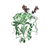
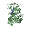
 HALOFERAX VOLCANII (archaea)
 X-RAY DIFFRACTION /
  Authors
 Citation
 Structure visualization
 Downloads & links
 Other downloads


 PDBj
PDBj


 Assembly
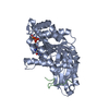


 Mass: 539.246 Da / Num. of mol.: 1 / Source method: obtained synthetically / Formula: C10H16N5O13P3S
 Mass: 18.015 Da / Num. of mol.: 99 / Source method: isolated from a natural source / Formula: H2O
 Sample preparation
 / Beamline: I03 / Wavelength: 0.9795
  Processing