[English] 日本語
Yorodumi
- PDB-4b0z: Crystal structure of S. pombe Rpn12 -

+
Open data


ID or keywords:

Loading...

-
Basic information

Entry
Database: PDB / ID: 4b0z
TitleCrystal structure of S. pombe Rpn12
Components26S PROTEASOME REGULATORY SUBUNIT RPN12
KeywordsPROTEIN BINDING / PROTEASOME UBITQUITIN
Function / homology
Function and homology information


Cross-presentation of soluble exogenous antigens (endosomes) / Regulation of ornithine decarboxylase (ODC) / : / UCH proteinases / Ub-specific processing proteases / chromatin => GO:0000785 / positive regulation of mitotic metaphase/anaphase transition / proteasome regulatory particle, lid subcomplex / nuclear periphery / proteasome-mediated ubiquitin-dependent protein catabolic process ...Cross-presentation of soluble exogenous antigens (endosomes) / Regulation of ornithine decarboxylase (ODC) / : / UCH proteinases / Ub-specific processing proteases / chromatin => GO:0000785 / positive regulation of mitotic metaphase/anaphase transition / proteasome regulatory particle, lid subcomplex / nuclear periphery / proteasome-mediated ubiquitin-dependent protein catabolic process / nucleus / cytosol
Similarity search - Function
Serine Threonine Protein Phosphatase 5, Tetratricopeptide repeat - #990 / 26S proteasome non-ATPase regulatory subunit Rpn12 / CSN8/PSMD8/EIF3K / CSN8/PSMD8/EIF3K family / Proteasome component (PCI) domain / PCI domain profile. / Serine Threonine Protein Phosphatase 5, Tetratricopeptide repeat / Alpha Horseshoe / Mainly Alpha
Similarity search - Domain/homology
NITRATE ION / MONOTHIOGLYCEROL / 26S proteasome regulatory subunit rpn12
Similarity search - Component
Biological speciesSCHIZOSACCHAROMYCES POMBE (fission yeast)
MethodX-RAY DIFFRACTION / SYNCHROTRON / SAD / Resolution: 1.585 Å
AuthorsBoehringer, J. / Riedinger, C. / Paraskevopoulos, K. / Johnson, E.O.D. / Lowe, E.D. / Khoudian, C. / Smith, D. / Noble, M.E.M. / Gordon, C. / Endicott, J.A.
CitationJournal: Biochem.J. / Year: 2012
Title: Structural and Functional Characterisation of Rpn12 Identifies Residues Required for Rpn10 Proteasome Incorporation.
Authors: Boehringer, J. / Riedinger, C. / Paraskevopoulos, K. / Johnson, E.O.D. / Lowe, E.D. / Khoudian, C. / Smith, D. / Noble, M.E.M. / Gordon, C. / Endicott, J.A.
History
DepositionJul 6, 2012Deposition site: PDBE / Processing site: PDBE
Revision 1.0Sep 12, 2012Provider: repository / Type: Initial release
Revision 1.1Nov 7, 2012Group: Database references

-
Structure visualization

Structure viewerMolecule:
MolmilJmol/JSmol

Downloads & links

-
Assembly

Deposited unit
A: 26S PROTEASOME REGULATORY SUBUNIT RPN12
B: 26S PROTEASOME REGULATORY SUBUNIT RPN12
hetero molecules


Theoretical massNumber of molelcules
Total (without water)53,19311
Polymers52,4522
Non-polymers7419
Water6,557364
1


  • Idetical with deposited unit
  • defined by author&software
TypeNameSymmetry operationNumber
identity operation1_555x,y,z1
Buried area2590 Å2
ΔGint-11.2 kcal/mol
Surface area23520 Å2
MethodPISA
Unit cell
Length a, b, c (Å)41.780, 91.380, 143.340
Angle α, β, γ (deg.)90.00, 90.00, 90.00
Int Tables number19
Space group name H-MP212121

-
Components

#1: Protein 26S PROTEASOME REGULATORY SUBUNIT RPN12 / RPN12


Mass: 26225.963 Da / Num. of mol.: 2 / Fragment: RESIDUES 1-224
Source method: isolated from a genetically manipulated source
Source: (gene. exp.) SCHIZOSACCHAROMYCES POMBE (fission yeast)
Plasmid: PGEX-6P-1 / Production host: ESCHERICHIA COLI (E. coli) / Strain (production host): BL21(DE3) / References: UniProt: P50524
#2: Chemical
ChemComp-NO3 / NITRATE ION


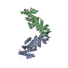
Mass: 62.005 Da / Num. of mol.: 4 / Source method: obtained synthetically / Formula: NO3
#3: Chemical ChemComp-SGM / MONOTHIOGLYCEROL


Mass: 108.159 Da / Num. of mol.: 2 / Source method: obtained synthetically / Formula: C3H8O2S
#4: Chemical ChemComp-GOL / GLYCEROL / GLYCERIN / PROPANE-1,2,3-TRIOL


Mass: 92.094 Da / Num. of mol.: 3 / Source method: obtained synthetically / Formula: C3H8O3
#5: Water ChemComp-HOH / water


Mass: 18.015 Da / Num. of mol.: 364 / Source method: isolated from a natural source / Formula: H2O
Nonpolymer detailsMONOTHIOGLYCEROL (SGM) IS BOUND TO CYS 65 BY A DISULPHIDE BRIDGE

-
Experimental details

-
Experiment

ExperimentMethod: X-RAY DIFFRACTION / Number of used crystals: 1

-
Sample preparation

CrystalDensity Matthews: 2.67 Å3/Da / Density % sol: 54 % / Description: NONE
Crystal growpH: 7.5
Details: 0.2 M NA NO3, 0.1 M BIS-TRIS PROPANE PH 7.5, 22.5% PEG3350

-
Data collection

DiffractionMean temperature: 100 K
Diffraction sourceSource: SYNCHROTRON / Site: ESRF / Beamline: ID14-2 / Wavelength: 0.9791
DetectorType: ADSC CCD / Detector: CCD / Date: Mar 17, 2008 / Details: TOROIDAL ZERODUR MIRROR
RadiationMonochromator: ASYMMETRIC LAUE 001 / Protocol: SINGLE WAVELENGTH / Monochromatic (M) / Laue (L): M / Scattering type: x-ray
Radiation wavelengthWavelength: 0.9791 Å / Relative weight: 1
ReflectionResolution: 1.59→42.34 Å / Num. obs: 74706 / % possible obs: 99.7 % / Observed criterion σ(I): 2 / Redundancy: 3.9 % / Biso Wilson estimate: 22.86 Å2 / Rmerge(I) obs: 0.05 / Net I/σ(I): 17.1
Reflection shellResolution: 1.59→1.67 Å / Redundancy: 3.9 % / Rmerge(I) obs: 0.5 / Mean I/σ(I) obs: 2.5 / % possible all: 99.7

-
Processing

Software
NameVersionClassification
PHENIX(PHENIX.REFINE)refinement
MOSFLMdata reduction
SCALAdata scaling
SHELXphasing
RefinementMethod to determine structure: SAD
Starting model: NONE

Resolution: 1.585→40.111 Å / SU ML: 0.17 / σ(F): 0 / Phase error: 18.43 / Stereochemistry target values: ML
RfactorNum. reflection% reflection
Rfree0.2248 3719 5.1 %
Rwork0.188 --
obs0.1898 73328 97.11 %
Solvent computationShrinkage radii: 0.9 Å / VDW probe radii: 1.11 Å / Solvent model: FLAT BULK SOLVENT MODEL / Bsol: 40.509 Å2 / ksol: 0.455 e/Å3
Displacement parametersBiso mean: 32.38 Å2
Baniso -1Baniso -2Baniso -3
1-0 Å20 Å20 Å2
2--0 Å20 Å2
3---0 Å2
Refinement stepCycle: LAST / Resolution: 1.585→40.111 Å
ProteinNucleic acidLigandSolventTotal
Num. atoms3606 0 46 364 4016
Refine LS restraints
Refine-IDTypeDev idealNumber
X-RAY DIFFRACTIONf_bond_d0.0153876
X-RAY DIFFRACTIONf_angle_d1.5255251
X-RAY DIFFRACTIONf_dihedral_angle_d13.5431464
X-RAY DIFFRACTIONf_chiral_restr0.081575
X-RAY DIFFRACTIONf_plane_restr0.007679
LS refinement shell
Resolution (Å)Rfactor RfreeNum. reflection RfreeRfactor RworkNum. reflection RworkRefine-ID% reflection obs (%)
1.585-1.64170.26643220.21026416X-RAY DIFFRACTION91
1.6417-1.70740.22483470.17536613X-RAY DIFFRACTION93
1.7074-1.78510.20293730.15196792X-RAY DIFFRACTION96
1.7851-1.87920.20053980.15226859X-RAY DIFFRACTION97
1.8792-1.99690.20983820.16036997X-RAY DIFFRACTION98
1.9969-2.15110.18873890.15677048X-RAY DIFFRACTION99
2.1511-2.36760.20893800.1487113X-RAY DIFFRACTION100
2.3676-2.71010.19333860.1557158X-RAY DIFFRACTION100
2.7101-3.41420.21813690.17657184X-RAY DIFFRACTION99
3.4142-40.12370.26253730.23857429X-RAY DIFFRACTION98

+
About Yorodumi

-
News

-
Feb 9, 2022. New format data for meta-information of EMDB entries

New format data for meta-information of EMDB entries

  • Version 3 of the EMDB header file is now the official format.
  • The previous official version 1.9 will be removed from the archive.

Related info.:EMDB header

External links:wwPDB to switch to version 3 of the EMDB data model

-
Aug 12, 2020. Covid-19 info

Covid-19 info

URL: https://pdbjlvh1.pdbj.org/emnavi/covid19.php

New page: Covid-19 featured information page in EM Navigator.

Related info.:Covid-19 info / Mar 5, 2020. Novel coronavirus structure data

+
Mar 5, 2020. Novel coronavirus structure data

Novel coronavirus structure data

Related info.:Yorodumi Speices / Aug 12, 2020. Covid-19 info

External links:COVID-19 featured content - PDBj / Molecule of the Month (242):Coronavirus Proteases

+
Jan 31, 2019. EMDB accession codes are about to change! (news from PDBe EMDB page)

EMDB accession codes are about to change! (news from PDBe EMDB page)

  • The allocation of 4 digits for EMDB accession codes will soon come to an end. Whilst these codes will remain in use, new EMDB accession codes will include an additional digit and will expand incrementally as the available range of codes is exhausted. The current 4-digit format prefixed with “EMD-” (i.e. EMD-XXXX) will advance to a 5-digit format (i.e. EMD-XXXXX), and so on. It is currently estimated that the 4-digit codes will be depleted around Spring 2019, at which point the 5-digit format will come into force.
  • The EM Navigator/Yorodumi systems omit the EMD- prefix.

Related info.:Q: What is EMD? / ID/Accession-code notation in Yorodumi/EM Navigator

External links:EMDB Accession Codes are Changing Soon! / Contact to PDBj

+
Jul 12, 2017. Major update of PDB

Major update of PDB

  • wwPDB released updated PDB data conforming to the new PDBx/mmCIF dictionary.
  • This is a major update changing the version number from 4 to 5, and with Remediation, in which all the entries are updated.
  • In this update, many items about electron microscopy experimental information are reorganized (e.g. em_software).
  • Now, EM Navigator and Yorodumi are based on the updated data.

External links:wwPDB Remediation / Enriched Model Files Conforming to OneDep Data Standards Now Available in the PDB FTP Archive

-
Yorodumi

Thousand views of thousand structures

  • Yorodumi is a browser for structure data from EMDB, PDB, SASBDB, etc.
  • This page is also the successor to EM Navigator detail page, and also detail information page/front-end page for Omokage search.
  • The word "yorodu" (or yorozu) is an old Japanese word meaning "ten thousand". "mi" (miru) is to see.

Related info.:EMDB / PDB / SASBDB / Comparison of 3 databanks / Yorodumi Search / Aug 31, 2016. New EM Navigator & Yorodumi / Yorodumi Papers / Jmol/JSmol / Function and homology information / Changes in new EM Navigator and Yorodumi

Read more