 Movie
Movie Controller

[English] 日本語
 Yorodumi
- PDB-4azq: Murine epidermal fatty acid-binding protein (FABP5) in complex wi... -
+ Open data

- Basic information
| Entry | Database: PDB / ID: 4azq | ||||||
|---|---|---|---|---|---|---|---|
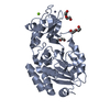
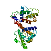
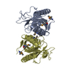
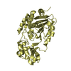
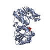
| Title | Murine epidermal fatty acid-binding protein (FABP5) in complex with the endocannabinoid 2-arachidonoylglycerol | ||||||
 Components Components | FATTY ACID-BINDING PROTEIN, EPIDERMAL | ||||||
 Keywords Keywords | LIPID BINDING PROTEIN / LIPID CARRIER PROTEIN / ENDOCANNABINOID / ANANDAMIDE / BETA-BARREL / BETA-CLAMSHELL / DOMAIN SWAPPING | ||||||
| Function / homology |  Function and homology information Function and homology informationSignaling by Retinoic Acid / regulation of prostaglandin biosynthetic process / regulation of retrograde trans-synaptic signaling by endocanabinoid / Triglyceride catabolism / lipid transport across blood-brain barrier / retrograde trans-synaptic signaling by endocannabinoid / positive regulation of peroxisome proliferator activated receptor signaling pathway / negative regulation of D-glucose transmembrane transport / regulation of sensory perception of pain / phosphatidylcholine biosynthetic process ...Signaling by Retinoic Acid / regulation of prostaglandin biosynthetic process / regulation of retrograde trans-synaptic signaling by endocanabinoid / Triglyceride catabolism / lipid transport across blood-brain barrier / retrograde trans-synaptic signaling by endocannabinoid / positive regulation of peroxisome proliferator activated receptor signaling pathway / negative regulation of D-glucose transmembrane transport / regulation of sensory perception of pain / phosphatidylcholine biosynthetic process / retinoic acid binding / long-chain fatty acid transmembrane transporter activity / fatty acid transport / postsynaptic cytosol / postsynaptic density, intracellular component / Neutrophil degranulation / fatty acid binding / lipid metabolic process / glucose metabolic process / glucose homeostasis / positive regulation of cold-induced thermogenesis / postsynaptic density / synapse / glutamatergic synapse / : / identical protein binding / nucleus / cytosol / cytoplasm Similarity search - Function | ||||||
| Biological species |  | ||||||
| Method |  X-RAY DIFFRACTION / SYNCHROTRON / MOLECULAR REPLACEMENT / Resolution: 2 Å X-RAY DIFFRACTION / SYNCHROTRON / MOLECULAR REPLACEMENT / Resolution: 2 Å | ||||||
 Authors Authors | Sanson, B. / Wang, T. / Sun, J. / Kaczocha, M. / Ojima, I. / Deutsch, D. / Li, H. | ||||||
 Citation Citation | Journal: Acta Crystallogr.,Sect.D / Year: 2014 Title: Crystallographic Study of Fabp5 as an Intracellular Endocannabinoid Transporter. Authors: Sanson, B. / Wang, T. / Sun, J. / Wang, L. / Kaczocha, M. / Ojima, I. / Deutsch, D. / Li, H. | ||||||
| History |
|
- Structure visualization
| Structure viewer | Molecule: MolmilJmol/JSmol |
|---|
- Downloads & links
-Download
| PDBx/mmCIF format | 4azq.cif.gz | 73.4 KB | Display | PDBx/mmCIF format |
|---|---|---|---|---|
| PDB format | pdb4azq.ent.gz | 53.6 KB | Display | PDB format |
| PDBx/mmJSON format | 4azq.json.gz | Tree view | PDBx/mmJSON format | |
| Others |  Other downloads Other downloads |
-Validation report
| Arichive directory | https://data.pdbj.org/pub/pdb/validation_reports/az/4azqftp://data.pdbj.org/pub/pdb/validation_reports/az/4azq | HTTPS FTP |
|---|
-Related structure data
| Related structure data | 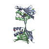 4azmC  4aznC  4azoC  4azpC 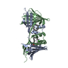 4azrC 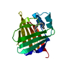 1b56S C: citing same article ( S: Starting model for refinement |
|---|---|
| Similar structure data |


-Links
 PDBj
PDBj


- Assembly
| Deposited unit | 
| ||||||||
|---|---|---|---|---|---|---|---|---|---|
| 1 | 
| ||||||||
| Unit cell |
| ||||||||
| Components on special symmetry positions |
|
-Components
| #1: Protein | Mass: 15454.736 Da / Num. of mol.: 1 Source method: isolated from a genetically manipulated source Source: (gene. exp.)  |
|---|---|
| #2: Chemical | ChemComp-G2A /   Mass: 386.609 Da / Num. of mol.: 1 / Source method: obtained synthetically / Formula: C23H46O4 Mass: 386.609 Da / Num. of mol.: 1 / Source method: obtained synthetically / Formula: C23H46O4 |
| #3: Chemical | ChemComp-CL /   Mass: 35.453 Da / Num. of mol.: 1 / Source method: obtained synthetically / Formula: Cl Mass: 35.453 Da / Num. of mol.: 1 / Source method: obtained synthetically / Formula: Cl |
| #4: Water | ChemComp-HOH /  Mass: 18.015 Da / Num. of mol.: 68 / Source method: isolated from a natural source / Formula: H2O Mass: 18.015 Da / Num. of mol.: 68 / Source method: isolated from a natural source / Formula: H2O |
| Sequence details | ADDITIONAL |
-Experimental details
-Experiment
| Experiment | Method: X-RAY DIFFRACTION / Number of used crystals: 1 |
|---|
- Sample preparation
| Crystal | Density Matthews: 2.4 Å3/Da / Density % sol: 49 % / Description: NONE |
|---|---|
| Crystal grow | Temperature: 293 K / Method: vapor diffusion, hanging drop / pH: 4.8 Details: 200 MM NACL, 50 MM NAAC PH 4.8, 25% PEG 3350 AND 5% MPD, VAPOR DIFFUSION, HANGING DROP, TEMPERATURE 293K. CRYSTALS SOAKED IN THE MOTHER LIQUOR SATURATED WITH 2-ARACHIDONOYLGLYCEROL AND ...Details: 200 MM NACL, 50 MM NAAC PH 4.8, 25% PEG 3350 AND 5% MPD, VAPOR DIFFUSION, HANGING DROP, TEMPERATURE 293K. CRYSTALS SOAKED IN THE MOTHER LIQUOR SATURATED WITH 2-ARACHIDONOYLGLYCEROL AND CONTAINING 25% GLYCEROL FOR CRYOPROTECTION. |
-Data collection
| Diffraction | Mean temperature: 100 K |
|---|---|
| Diffraction source | Source: SYNCHROTRON / Site: NSLS  / Beamline: X29A / Wavelength: 1.075 / Beamline: X29A / Wavelength: 1.075 |
| Detector | Type: ADSC QUANTUM 315r / Detector: CCD / Date: Sep 14, 2010 |
| Radiation | Monochromator: SI(111) / Protocol: SINGLE WAVELENGTH / Monochromatic (M) / Laue (L): M / Scattering type: x-ray |
| Radiation wavelength | Wavelength: 1.075 Å / Relative weight: 1 |
| Reflection | Resolution: 2→20 Å / Num. obs: 10920 / % possible obs: 99.6 % / Observed criterion σ(I): -3 / Redundancy: 11.4 % / Biso Wilson estimate: 51.5 Å2 / Rmerge(I) obs: 0.08 / Net I/σ(I): 18.5 |
| Reflection shell | Resolution: 2→2.1 Å / Redundancy: 11.7 % / Rmerge(I) obs: 0.52 / Mean I/σ(I) obs: 4.9 / % possible all: 100 |
- Processing
| Software |
| ||||||||||||||||||||||||||||||||||||||||||||||||||||||||||||||||||||||||||||||||||||||||||||||||||||||||||||||||||||||||||||||||||||||||||||||||||||||||||||||||||||||||||||||||||||||
|---|---|---|---|---|---|---|---|---|---|---|---|---|---|---|---|---|---|---|---|---|---|---|---|---|---|---|---|---|---|---|---|---|---|---|---|---|---|---|---|---|---|---|---|---|---|---|---|---|---|---|---|---|---|---|---|---|---|---|---|---|---|---|---|---|---|---|---|---|---|---|---|---|---|---|---|---|---|---|---|---|---|---|---|---|---|---|---|---|---|---|---|---|---|---|---|---|---|---|---|---|---|---|---|---|---|---|---|---|---|---|---|---|---|---|---|---|---|---|---|---|---|---|---|---|---|---|---|---|---|---|---|---|---|---|---|---|---|---|---|---|---|---|---|---|---|---|---|---|---|---|---|---|---|---|---|---|---|---|---|---|---|---|---|---|---|---|---|---|---|---|---|---|---|---|---|---|---|---|---|---|---|---|---|
| Refinement | Method to determine structure: MOLECULAR REPLACEMENT Starting model: PDB ENTRY 1B56 Resolution: 2→20 Å / Cor.coef. Fo:Fc: 0.95 / Cor.coef. Fo:Fc free: 0.94 / SU B: 10.907 / SU ML: 0.137 / Cross valid method: THROUGHOUT / ESU R: 0.201 / ESU R Free: 0.176 / Stereochemistry target values: MAXIMUM LIKELIHOOD / Details: HYDROGENS HAVE BEEN ADDED IN THE RIDING POSITIONS.
| ||||||||||||||||||||||||||||||||||||||||||||||||||||||||||||||||||||||||||||||||||||||||||||||||||||||||||||||||||||||||||||||||||||||||||||||||||||||||||||||||||||||||||||||||||||||
| Solvent computation | Ion probe radii: 0.8 Å / Shrinkage radii: 0.8 Å / VDW probe radii: 1.4 Å / Solvent model: MASK | ||||||||||||||||||||||||||||||||||||||||||||||||||||||||||||||||||||||||||||||||||||||||||||||||||||||||||||||||||||||||||||||||||||||||||||||||||||||||||||||||||||||||||||||||||||||
| Displacement parameters | Biso mean: 44.061 Å2
| ||||||||||||||||||||||||||||||||||||||||||||||||||||||||||||||||||||||||||||||||||||||||||||||||||||||||||||||||||||||||||||||||||||||||||||||||||||||||||||||||||||||||||||||||||||||
| Refinement step | Cycle: LAST / Resolution: 2→20 Å
| ||||||||||||||||||||||||||||||||||||||||||||||||||||||||||||||||||||||||||||||||||||||||||||||||||||||||||||||||||||||||||||||||||||||||||||||||||||||||||||||||||||||||||||||||||||||
| Refine LS restraints |
|