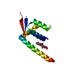
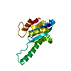
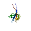
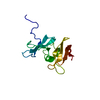
[English] 日本語
Yorodumi
- PDB-4aya: Crystal structure of ID2 HLH homodimer at 2.1A resolution -

+
Open data


ID or keywords:

Loading...

-
Basic information

Entry
Database: PDB / ID: 4aya
TitleCrystal structure of ID2 HLH homodimer at 2.1A resolution
ComponentsDNA-BINDING PROTEIN INHIBITOR ID-2
KeywordsCELL CYCLE
Function / homology
Function and homology information


: / endodermal digestive tract morphogenesis / euchromatin => GO:0000791 / bundle of His development / regulation of cell cycle => GO:0051726 / enucleate erythrocyte differentiation / negative regulation of B cell differentiation / RNA polymerase II-specific DNA-binding transcription factor binding => GO:0061629 / epithelial cell differentiation involved in mammary gland alveolus development / cell morphogenesis involved in neuron differentiation ...: / endodermal digestive tract morphogenesis / euchromatin => GO:0000791 / bundle of His development / regulation of cell cycle => GO:0051726 / enucleate erythrocyte differentiation / negative regulation of B cell differentiation / RNA polymerase II-specific DNA-binding transcription factor binding => GO:0061629 / epithelial cell differentiation involved in mammary gland alveolus development / cell morphogenesis involved in neuron differentiation / negative regulation of core promoter binding / negative regulation of muscle cell differentiation / thigmotaxis / natural killer cell differentiation / membranous septum morphogenesis / embryonic digestive tract morphogenesis / neuron fate commitment / positive regulation of macrophage differentiation / oligodendrocyte development / regulation of neural precursor cell proliferation / negative regulation of oligodendrocyte differentiation / olfactory bulb development / positive regulation of astrocyte differentiation / Peyer's patch development / positive regulation of blood pressure / metanephros development / NGF-stimulated transcription / transcription regulator inhibitor activity / mammary gland epithelial cell proliferation / regulation of neuron differentiation / cellular response to lithium ion / entrainment of circadian clock by photoperiod / regulation of G1/S transition of mitotic cell cycle / locomotor rhythm / regulation of lipid metabolic process / negative regulation of neuron differentiation / positive regulation of fat cell differentiation / mammary gland alveolus development / negative regulation of osteoblast differentiation / adipose tissue development / cell maturation / adult locomotory behavior / positive regulation of erythrocyte differentiation / positive regulation of smooth muscle cell proliferation / circadian regulation of gene expression / regulation of circadian rhythm / negative regulation of DNA-binding transcription factor activity / cellular senescence / transmembrane transporter binding / cell differentiation / protein dimerization activity / negative regulation of gene expression / negative regulation of DNA-templated transcription / positive regulation of gene expression / positive regulation of DNA-templated transcription / negative regulation of transcription by RNA polymerase II / protein-containing complex / nucleus / cytosol / cytoplasm
Similarity search - Function
DNA-binding protein inhibitor / Helix-loop-helix DNA-binding domain / MYOD Basic-Helix-Loop-Helix Domain, subunit B / Helix-loop-helix DNA-binding domain / Helix-loop-helix DNA-binding domain superfamily / helix loop helix domain / Myc-type, basic helix-loop-helix (bHLH) domain / Myc-type, basic helix-loop-helix (bHLH) domain profile. / Few Secondary Structures / Irregular
Similarity search - Domain/homology
ACETATE ION / : / DNA-binding protein inhibitor ID-2
Similarity search - Component
Biological speciesHOMO SAPIENS (human)
MethodX-RAY DIFFRACTION / SYNCHROTRON / MOLECULAR REPLACEMENT / Resolution: 2.103 Å
AuthorsWong, M.V. / Jiang, S. / Palasingam, P. / Kolatkar, P.R.
CitationJournal: Plos One / Year: 2012
Title: A Divalent Ion is Crucial in the Structure and Dominant-Negative Function of Id Proteins, a Class of Helix-Loop-Helix Transcription Regulators.
Authors: Wong, M.V. / Jiang, S. / Palasingam, P. / Kolatkar, P.R.
History
DepositionJun 19, 2012Deposition site: PDBE / Processing site: PDBE
Revision 1.0Nov 14, 2012Provider: repository / Type: Initial release
Revision 1.1May 1, 2024Group: Data collection / Database references ...Data collection / Database references / Derived calculations / Other / Refinement description
Category: chem_comp_atom / chem_comp_bond ...chem_comp_atom / chem_comp_bond / database_2 / pdbx_database_status / pdbx_initial_refinement_model / pdbx_struct_conn_angle / struct_conn / struct_site
Item: _database_2.pdbx_DOI / _database_2.pdbx_database_accession ..._database_2.pdbx_DOI / _database_2.pdbx_database_accession / _pdbx_database_status.status_code_sf / _pdbx_struct_conn_angle.ptnr1_auth_comp_id / _pdbx_struct_conn_angle.ptnr1_auth_seq_id / _pdbx_struct_conn_angle.ptnr1_label_asym_id / _pdbx_struct_conn_angle.ptnr1_label_atom_id / _pdbx_struct_conn_angle.ptnr1_label_comp_id / _pdbx_struct_conn_angle.ptnr1_label_seq_id / _pdbx_struct_conn_angle.ptnr3_auth_comp_id / _pdbx_struct_conn_angle.ptnr3_auth_seq_id / _pdbx_struct_conn_angle.ptnr3_label_asym_id / _pdbx_struct_conn_angle.ptnr3_label_atom_id / _pdbx_struct_conn_angle.ptnr3_label_comp_id / _pdbx_struct_conn_angle.ptnr3_label_seq_id / _pdbx_struct_conn_angle.value / _struct_conn.pdbx_dist_value / _struct_conn.ptnr1_auth_comp_id / _struct_conn.ptnr1_auth_seq_id / _struct_conn.ptnr1_label_asym_id / _struct_conn.ptnr1_label_atom_id / _struct_conn.ptnr1_label_comp_id / _struct_conn.ptnr1_label_seq_id / _struct_conn.ptnr2_auth_comp_id / _struct_conn.ptnr2_auth_seq_id / _struct_conn.ptnr2_label_asym_id / _struct_conn.ptnr2_label_atom_id / _struct_conn.ptnr2_label_comp_id / _struct_conn.ptnr2_label_seq_id / _struct_site.pdbx_auth_asym_id / _struct_site.pdbx_auth_comp_id / _struct_site.pdbx_auth_seq_id

-
Structure visualization

Structure viewerMolecule:
MolmilJmol/JSmol

Downloads & links

-
Assembly

Deposited unit
A: DNA-BINDING PROTEIN INHIBITOR ID-2
B: DNA-BINDING PROTEIN INHIBITOR ID-2
hetero molecules


Theoretical massNumber of molelcules
Total (without water)21,8265
Polymers21,6892
Non-polymers1373
Water43224
1


  • Idetical with deposited unit
  • defined by author&software
TypeNameSymmetry operationNumber
identity operation1_555x,y,z1
Buried area2350 Å2
ΔGint-30.1 kcal/mol
Surface area7350 Å2
MethodPISA
Unit cell
Length a, b, c (Å)51.622, 51.622, 111.474
Angle α, β, γ (deg.)90.00, 90.00, 120.00
Int Tables number154
Space group name H-MP3221

-
Components

#1: Protein DNA-BINDING PROTEIN INHIBITOR ID-2 / CLASS D BASIC HELIX-LOOP-HELIX PROTEIN 26 / CLASS B BASIC HELIX-LOOP-HELIX PROTEIN 26 / BHLHB26 / ...CLASS D BASIC HELIX-LOOP-HELIX PROTEIN 26 / CLASS B BASIC HELIX-LOOP-HELIX PROTEIN 26 / BHLHB26 / INHIBITOR OF DNA BINDING 2


Mass: 10844.563 Da / Num. of mol.: 2 / Fragment: HELIX-LOOP-HELIX DOMAIN, RESIDUES 1-82
Source method: isolated from a genetically manipulated source
Source: (gene. exp.) HOMO SAPIENS (human) / Plasmid: PDEST-565 / Production host: ESCHERICHIA COLI (E. coli) / Strain (production host): BL21(DE3) / References: UniProt: Q02363
#2: Chemical ChemComp-K / POTASSIUM ION


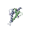
Mass: 39.098 Da / Num. of mol.: 2 / Source method: obtained synthetically / Formula: K
#3: Chemical ChemComp-ACT / ACETATE ION / Acetate


Mass: 59.044 Da / Num. of mol.: 1 / Source method: obtained synthetically / Formula: C2H3O2
#4: Water ChemComp-HOH / water / Water


Mass: 18.015 Da / Num. of mol.: 24 / Source method: isolated from a natural source / Formula: H2O
Sequence detailsFIRST RESIDUE IN SEQUENCE AFTER TEV CLEAVAGE IS A GLYCINE. ID2 SEQUENCE NUMBERING STARTS FROM ...FIRST RESIDUE IN SEQUENCE AFTER TEV CLEAVAGE IS A GLYCINE. ID2 SEQUENCE NUMBERING STARTS FROM SECOND RESIDUE, METHIONINE. RESIDUE 83 ONWARDS CONTAINS THE C-TERMINAL STABILIZER POLYPEPTIDE.

-
Experimental details

-
Experiment

ExperimentMethod: X-RAY DIFFRACTION / Number of used crystals: 1

-
Sample preparation

CrystalDensity Matthews: 1.98 Å3/Da / Density % sol: 37.8 %
Crystal growpH: 6.5
Details: PROTEIN WAS CRYSTALLIZED FROM 0.1 M MES PH6.5, 2.0 M POTASSIUM ACETATE

-
Data collection

DiffractionMean temperature: 103 K
Diffraction sourceSource: SYNCHROTRON / Site: NSLS / Beamline: X29A / Wavelength: 1.0809
DetectorType: ADSC CCD / Detector: CCD / Date: Jun 29, 2009
RadiationProtocol: SINGLE WAVELENGTH / Monochromatic (M) / Laue (L): M / Scattering type: x-ray
Radiation wavelengthWavelength: 1.0809 Å / Relative weight: 1
ReflectionResolution: 2.1→50 Å / Num. obs: 10569 / % possible obs: 100 % / Observed criterion σ(I): 0 / Redundancy: 11.9 % / Biso Wilson estimate: 41.98 Å2 / Rmerge(I) obs: 0.06 / Net I/σ(I): 40.6
Reflection shellResolution: 2.1→2.18 Å / Redundancy: 10.2 % / Rmerge(I) obs: 0.42 / Mean I/σ(I) obs: 4.2 / % possible all: 100

-
Processing

Software
NameVersionClassification
PHENIX(PHENIX.REFINE)refinement
HKL-2000data reduction
SCALEPACKdata scaling
PHENIXphasing
RefinementMethod to determine structure: MOLECULAR REPLACEMENT
Starting model: INTERNAL SELENOMETHIONINE-SUBSTITUTED ID2 STRUCTURE AT 3.0A

Resolution: 2.103→44.706 Å / SU ML: 0.23 / σ(F): 0.65 / Phase error: 27.31 / Stereochemistry target values: ML
Details: RESIDUES 1-29 ARE MISSING IN CHAIN A. RESIDUES 1-34 ARE MISSING IN CHAIN B.
RfactorNum. reflection% reflection
Rfree0.2497 987 9.8 %
Rwork0.2246 --
obs0.2271 10026 95.2 %
Solvent computationShrinkage radii: 0.9 Å / VDW probe radii: 1.11 Å / Solvent model: FLAT BULK SOLVENT MODEL / Bsol: 0 Å2 / ksol: 0 e/Å3
Displacement parametersBiso mean: 54.4 Å2
Refinement stepCycle: LAST / Resolution: 2.103→44.706 Å
ProteinNucleic acidLigandSolventTotal
Num. atoms870 0 6 24 900
Refine LS restraints
Refine-IDTypeDev idealNumber
X-RAY DIFFRACTIONf_bond_d0.007888
X-RAY DIFFRACTIONf_angle_d1.0711198
X-RAY DIFFRACTIONf_dihedral_angle_d13.029346
X-RAY DIFFRACTIONf_chiral_restr0.059141
X-RAY DIFFRACTIONf_plane_restr0.004149
LS refinement shell
Resolution (Å)Rfactor RfreeNum. reflection RfreeRfactor RworkNum. reflection RworkRefine-ID% reflection obs (%)
2.1031-2.21390.30491290.25131147X-RAY DIFFRACTION88
2.2139-2.35260.31241360.23511228X-RAY DIFFRACTION91
2.3526-2.53430.30811330.23571229X-RAY DIFFRACTION94
2.5343-2.78930.27751400.25851300X-RAY DIFFRACTION97
2.7893-3.19280.32681480.26131335X-RAY DIFFRACTION98
3.1928-4.02220.24791480.20961356X-RAY DIFFRACTION100
4.0222-44.7160.19681530.20911444X-RAY DIFFRACTION99

+
About Yorodumi

-
News

-
Feb 9, 2022. New format data for meta-information of EMDB entries

New format data for meta-information of EMDB entries

  • Version 3 of the EMDB header file is now the official format.
  • The previous official version 1.9 will be removed from the archive.

Related info.:EMDB header

External links:wwPDB to switch to version 3 of the EMDB data model

-
Aug 12, 2020. Covid-19 info

Covid-19 info

URL: https://pdbj.org/emnavi/covid19.php

New page: Covid-19 featured information page in EM Navigator.

Related info.:Covid-19 info / Mar 5, 2020. Novel coronavirus structure data

+
Mar 5, 2020. Novel coronavirus structure data

Novel coronavirus structure data

Related info.:Yorodumi Speices / Aug 12, 2020. Covid-19 info

External links:COVID-19 featured content - PDBj / Molecule of the Month (242):Coronavirus Proteases

+
Jan 31, 2019. EMDB accession codes are about to change! (news from PDBe EMDB page)

EMDB accession codes are about to change! (news from PDBe EMDB page)

  • The allocation of 4 digits for EMDB accession codes will soon come to an end. Whilst these codes will remain in use, new EMDB accession codes will include an additional digit and will expand incrementally as the available range of codes is exhausted. The current 4-digit format prefixed with “EMD-” (i.e. EMD-XXXX) will advance to a 5-digit format (i.e. EMD-XXXXX), and so on. It is currently estimated that the 4-digit codes will be depleted around Spring 2019, at which point the 5-digit format will come into force.
  • The EM Navigator/Yorodumi systems omit the EMD- prefix.

Related info.:Q: What is EMD? / ID/Accession-code notation in Yorodumi/EM Navigator

External links:EMDB Accession Codes are Changing Soon! / Contact to PDBj

+
Jul 12, 2017. Major update of PDB

Major update of PDB

  • wwPDB released updated PDB data conforming to the new PDBx/mmCIF dictionary.
  • This is a major update changing the version number from 4 to 5, and with Remediation, in which all the entries are updated.
  • In this update, many items about electron microscopy experimental information are reorganized (e.g. em_software).
  • Now, EM Navigator and Yorodumi are based on the updated data.

External links:wwPDB Remediation / Enriched Model Files Conforming to OneDep Data Standards Now Available in the PDB FTP Archive

-
Yorodumi

Thousand views of thousand structures

  • Yorodumi is a browser for structure data from EMDB, PDB, SASBDB, etc.
  • This page is also the successor to EM Navigator detail page, and also detail information page/front-end page for Omokage search.
  • The word "yorodu" (or yorozu) is an old Japanese word meaning "ten thousand". "mi" (miru) is to see.

Related info.:EMDB / PDB / SASBDB / Comparison of 3 databanks / Yorodumi Search / Aug 31, 2016. New EM Navigator & Yorodumi / Yorodumi Papers / Jmol/JSmol / Function and homology information / Changes in new EM Navigator and Yorodumi

Read more