 Movie
Movie Controller

[English] 日本語
 Yorodumi
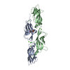
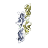
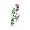
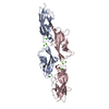
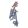
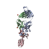
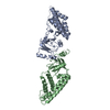
- PDB-4axw: CRYSTAL STRUCTURE OF MOUSE CADHERIN-23 EC1-2 AND PROTOCADHERIN-15... -
+ Open data

- Basic information
| Entry | Database: PDB / ID: 4axw | ||||||
|---|---|---|---|---|---|---|---|
| Title | CRYSTAL STRUCTURE OF MOUSE CADHERIN-23 EC1-2 AND PROTOCADHERIN-15 EC1- 2, FORM I 2.2A. | ||||||
 Components Components |
| ||||||
 Keywords Keywords | CELL ADHESION / HEARING / DEAFNESS / CADHERIN / CDH23 / PCDH15 / HETEROPHILIC | ||||||
| Function / homology |  Function and homology information Function and homology informationdetection of mechanical stimulus involved in equilibrioception / cochlear hair cell ribbon synapse / equilibrioception / righting reflex / sensory perception of light stimulus / inner ear receptor cell stereocilium organization / stereocilium tip / photoreceptor ribbon synapse / inner ear auditory receptor cell differentiation / kinocilium ...detection of mechanical stimulus involved in equilibrioception / cochlear hair cell ribbon synapse / equilibrioception / righting reflex / sensory perception of light stimulus / inner ear receptor cell stereocilium organization / stereocilium tip / photoreceptor ribbon synapse / inner ear auditory receptor cell differentiation / kinocilium / stereocilium bundle / detection of mechanical stimulus involved in sensory perception of sound / calcium-dependent cell-cell adhesion / photoreceptor cell maintenance / catenin complex / stereocilium / non-motile cilium assembly / auditory receptor cell stereocilium organization / adult walking behavior / inner ear morphogenesis / inner ear development / homophilic cell-cell adhesion / startle response / actin filament bundle assembly / cochlea development / photoreceptor outer segment / regulation of cytosolic calcium ion concentration / photoreceptor inner segment / visual perception / cell adhesion molecule binding / morphogenesis of an epithelium / multicellular organism growth / actin filament organization / sensory perception of sound / locomotory behavior / response to calcium ion / beta-catenin binding / neuron projection development / apical part of cell / calcium ion transport / cell migration / cell adhesion / cadherin binding / calcium ion binding / centrosome / synapse / : / membrane / plasma membrane / cytoplasm Similarity search - Function | ||||||
| Biological species |  | ||||||
| Method |  X-RAY DIFFRACTION / SYNCHROTRON / MOLECULAR REPLACEMENT / Resolution: 2.23 Å X-RAY DIFFRACTION / SYNCHROTRON / MOLECULAR REPLACEMENT / Resolution: 2.23 Å | ||||||
 Authors Authors | Sotomayor, M. / Weihofen, W. / Gaudet, R. / Corey, D.P. | ||||||
 Citation Citation | Journal: Nature / Year: 2012 Title: Structure of a Force-Conveying Cadherin Bond Essential for Inner-Ear Mechanotransduction Authors: Sotomayor, M. / Weihofen, W. / Gaudet, R. / Corey, D.P. | ||||||
| History |
|
- Structure visualization
| Structure viewer | Molecule: MolmilJmol/JSmol |
|---|
- Downloads & links
-Download
| PDBx/mmCIF format | 4axw.cif.gz | 207.3 KB | Display | PDBx/mmCIF format |
|---|---|---|---|---|
| PDB format | pdb4axw.ent.gz | 164.9 KB | Display | PDB format |
| PDBx/mmJSON format | 4axw.json.gz | Tree view | PDBx/mmJSON format | |
| Others |  Other downloads Other downloads |
-Validation report
| Arichive directory | https://data.pdbj.org/pub/pdb/validation_reports/ax/4axwftp://data.pdbj.org/pub/pdb/validation_reports/ax/4axw | HTTPS FTP |
|---|
-Related structure data
| Related structure data |  4apxC  4aq8C  4aqaC  4aqeC  2whvS S: Starting model for refinement C: citing same article ( |
|---|---|
| Similar structure data |


-Links
 PDBj
PDBj
- Assembly
| Deposited unit | 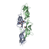
| ||||||||
|---|---|---|---|---|---|---|---|---|---|
| 1 |
| ||||||||
| Unit cell |
| ||||||||
| Components on special symmetry positions |
|
-Components
-Protein , 2 types, 2 molecules AB
| #1: Protein | Mass: 23856.436 Da / Num. of mol.: 1 / Fragment: EC1-2, RESIDUES 24-228 Source method: isolated from a genetically manipulated source Source: (gene. exp.)  |
|---|---|
| #2: Protein | Mass: 27502.619 Da / Num. of mol.: 1 / Fragment: EC1-2, RESIDUES 27-259 Source method: isolated from a genetically manipulated source Source: (gene. exp.) |
-Non-polymers , 7 types, 360 molecules 


| #3: Chemical | ChemComp-CA /  Mass: 40.078 Da / Num. of mol.: 7 / Source method: obtained synthetically / Formula: Ca Mass: 40.078 Da / Num. of mol.: 7 / Source method: obtained synthetically / Formula: Ca#4: Chemical | ChemComp-MES / |  Mass: 195.237 Da / Num. of mol.: 1 / Source method: obtained synthetically / Formula: C6H13NO4S / Comment: pH buffer*YM Mass: 195.237 Da / Num. of mol.: 1 / Source method: obtained synthetically / Formula: C6H13NO4S / Comment: pH buffer*YM#5: Chemical | ChemComp-PG4 / |  Mass: 194.226 Da / Num. of mol.: 1 / Source method: obtained synthetically / Formula: C8H18O5 / Comment: precipitant*YM Mass: 194.226 Da / Num. of mol.: 1 / Source method: obtained synthetically / Formula: C8H18O5 / Comment: precipitant*YM#6: Chemical | ChemComp-CL / |  Mass: 35.453 Da / Num. of mol.: 1 / Source method: obtained synthetically / Formula: Cl Mass: 35.453 Da / Num. of mol.: 1 / Source method: obtained synthetically / Formula: Cl#7: Chemical | ChemComp-GOL / |  Mass: 92.094 Da / Num. of mol.: 1 / Source method: obtained synthetically / Formula: C3H8O3 Mass: 92.094 Da / Num. of mol.: 1 / Source method: obtained synthetically / Formula: C3H8O3#8: Chemical | ChemComp-K / |  Mass: 39.098 Da / Num. of mol.: 1 / Source method: obtained synthetically / Formula: K Mass: 39.098 Da / Num. of mol.: 1 / Source method: obtained synthetically / Formula: K#9: Water | ChemComp-HOH / | Mass: 18.015 Da / Num. of mol.: 348 / Source method: isolated from a natural source / Formula: H2O |
|---|
-Details
| Has protein modification | Y |
|---|
-Experimental details
-Experiment
| Experiment | Method: X-RAY DIFFRACTION / Number of used crystals: 1 |
|---|
- Sample preparation
| Crystal | Density Matthews: 2.94 Å3/Da / Density % sol: 58.24 % / Description: NONE |
|---|---|
| Crystal grow | pH: 6.5 Details: 0.1 M MES PH 6.5, 15% W/V PEG550MME, 25% GLYCEROL, 0.2M POTASSIUM CHLORIDE |
-Data collection
| Diffraction | Mean temperature: 100 K |
|---|---|
| Diffraction source | Source: SYNCHROTRON / Site: ALS  / Beamline: 4.2.2 / Wavelength: 1.138 / Beamline: 4.2.2 / Wavelength: 1.138 |
| Detector | Type: E. WESTBROOK NOIR-1 MBC SYSTEM / Detector: CCD / Date: Dec 21, 2010 |
| Radiation | Monochromator: ROSENBAUM-ROCK SI(111) SAGITALLY FOCUSED / Protocol: SINGLE WAVELENGTH / Monochromatic (M) / Laue (L): M / Scattering type: x-ray |
| Radiation wavelength | Wavelength: 1.138 Å / Relative weight: 1 |
| Reflection | Resolution: 2.23→50 Å / Num. obs: 27442 / % possible obs: 96.2 % / Observed criterion σ(I): 2 / Redundancy: 3.4 % / Rmerge(I) obs: 0.11 / Net I/σ(I): 10.65 |
| Reflection shell | Resolution: 2.23→2.27 Å / Redundancy: 2.9 % / Rmerge(I) obs: 0.39 / Mean I/σ(I) obs: 2.69 / % possible all: 87.5 |
- Processing
| Software |
| ||||||||||||||||||||||||||||||||||||||||||||||||||||||||||||||||||||||||||||||||||||||||||||||||||||||||||||||||||||||||||||||||||||||||||||||||||||||||||||||||||||||||||||||||||||||
|---|---|---|---|---|---|---|---|---|---|---|---|---|---|---|---|---|---|---|---|---|---|---|---|---|---|---|---|---|---|---|---|---|---|---|---|---|---|---|---|---|---|---|---|---|---|---|---|---|---|---|---|---|---|---|---|---|---|---|---|---|---|---|---|---|---|---|---|---|---|---|---|---|---|---|---|---|---|---|---|---|---|---|---|---|---|---|---|---|---|---|---|---|---|---|---|---|---|---|---|---|---|---|---|---|---|---|---|---|---|---|---|---|---|---|---|---|---|---|---|---|---|---|---|---|---|---|---|---|---|---|---|---|---|---|---|---|---|---|---|---|---|---|---|---|---|---|---|---|---|---|---|---|---|---|---|---|---|---|---|---|---|---|---|---|---|---|---|---|---|---|---|---|---|---|---|---|---|---|---|---|---|---|---|
| Refinement | Method to determine structure: MOLECULAR REPLACEMENT Starting model: PDB ENTRY 2WHV Resolution: 2.23→41.08 Å / Cor.coef. Fo:Fc: 0.955 / Cor.coef. Fo:Fc free: 0.921 / SU B: 11.598 / SU ML: 0.133 / Cross valid method: THROUGHOUT / ESU R: 0.249 / ESU R Free: 0.212 / Stereochemistry target values: MAXIMUM LIKELIHOOD Details: HYDROGENS HAVE BEEN ADDED IN THE RIDING POSITIONS. U VALUES WITH TLS ADDED
| ||||||||||||||||||||||||||||||||||||||||||||||||||||||||||||||||||||||||||||||||||||||||||||||||||||||||||||||||||||||||||||||||||||||||||||||||||||||||||||||||||||||||||||||||||||||
| Solvent computation | Ion probe radii: 0.8 Å / Shrinkage radii: 0.8 Å / VDW probe radii: 1.4 Å / Solvent model: MASK | ||||||||||||||||||||||||||||||||||||||||||||||||||||||||||||||||||||||||||||||||||||||||||||||||||||||||||||||||||||||||||||||||||||||||||||||||||||||||||||||||||||||||||||||||||||||
| Displacement parameters | Biso mean: 23.536 Å2
| ||||||||||||||||||||||||||||||||||||||||||||||||||||||||||||||||||||||||||||||||||||||||||||||||||||||||||||||||||||||||||||||||||||||||||||||||||||||||||||||||||||||||||||||||||||||
| Refinement step | Cycle: LAST / Resolution: 2.23→41.08 Å
| ||||||||||||||||||||||||||||||||||||||||||||||||||||||||||||||||||||||||||||||||||||||||||||||||||||||||||||||||||||||||||||||||||||||||||||||||||||||||||||||||||||||||||||||||||||||
| Refine LS restraints |
|