Mass: 18.015 Da / Num. of mol.: 366 / Source method: isolated from a natural source / Formula: H2O
Sequence details
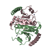
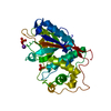
N-TERMINAL TRUNCATION, START AT RESIDUE 17. METHIONINE AND GLYCINE FROM EXPRESSION VECTOR AT N- ...N-TERMINAL TRUNCATION, START AT RESIDUE 17. METHIONINE AND GLYCINE FROM EXPRESSION VECTOR AT N-TERMINUS, C-TERMINAL HIS-TAG.
-
Experimental details
-
Experiment
Experiment
Method: X-RAY DIFFRACTION / Number of used crystals: 1
-
Sample preparation
Crystal
Density Matthews: 2.12 Å3/Da / Density % sol: 42.05 % / Description: NONE
Crystal grow
Method: vapor diffusion, hanging drop / pH: 4.5 Details: PROTEIN IN 150 MM NACL, 50 MM TRIS.HCL PH 8.0 AT 8 MG/ML. HANGING DROP VAPOUR DIFFUSION, 1/1 UL DROPS OVER 1ML OF PRECIPITANT: 100 MM SODIUM ACETATE PH 4.5, 35 % (W/V) PEG 6000, 200 MM MGCL2. ...Details: PROTEIN IN 150 MM NACL, 50 MM TRIS.HCL PH 8.0 AT 8 MG/ML. HANGING DROP VAPOUR DIFFUSION, 1/1 UL DROPS OVER 1ML OF PRECIPITANT: 100 MM SODIUM ACETATE PH 4.5, 35 % (W/V) PEG 6000, 200 MM MGCL2. CRYPROTECTED WITH WELL SOLUTION SUPPLEMENTED WITH 20 % (V/V) PEG 300
Resolution: 1→36.503 Å / SU ML: 0.05 / σ(F): 1.34 / Phase error: 11.83 / Stereochemistry target values: ML Details: LOOP BETWEEN RESIDUES 56 AND 64 IN CHAIN A MODELLED IN TWO CONFORMATIONS WITH 0.55 AND 0.45 OCCUPANCY.
Rfactor
Num. reflection
% reflection
Rfree
0.147
7431
5 %
Rwork
0.1357
-
-
obs
0.1363
148298
97.76 %
Solvent computation
Shrinkage radii: 0.6 Å / VDW probe radii: 0.9 Å / Solvent model: FLAT BULK SOLVENT MODEL / Bsol: 42.675 Å2 / ksol: 0.431 e/Å3
Displacement parameters
Biso mean: 14.74 Å2
Baniso -1
Baniso -2
Baniso -3
1-
1.233 Å2
0 Å2
-0.5928 Å2
2-
-
1.9062 Å2
0 Å2
3-
-
-
-3.1392 Å2
Refinement step
Cycle: LAST / Resolution: 1→36.503 Å
Protein
Nucleic acid
Ligand
Solvent
Total
Num. atoms
2100
0
1
366
2467
Refine LS restraints
Refine-ID
Type
Dev ideal
Number
X-RAY DIFFRACTION
f_bond_d
0.014
2354
X-RAY DIFFRACTION
f_angle_d
1.601
3237
X-RAY DIFFRACTION
f_dihedral_angle_d
12.433
901
X-RAY DIFFRACTION
f_chiral_restr
0.098
369
X-RAY DIFFRACTION
f_plane_restr
0.011
415
LS refinement shell
Resolution (Å)
Rfactor Rfree
Num. reflection Rfree
Rfactor Rwork
Num. reflection Rwork
Refine-ID
% reflection obs (%)
1-1.0114
0.2427
252
0.2336
4723
X-RAY DIFFRACTION
99
1.0114-1.0233
0.2204
240
0.2174
4774
X-RAY DIFFRACTION
99
1.0233-1.0358
0.2137
246
0.1922
4749
X-RAY DIFFRACTION
99
1.0358-1.0489
0.2088
255
0.1851
4735
X-RAY DIFFRACTION
99
1.0489-1.0627
0.1862
236
0.1695
4805
X-RAY DIFFRACTION
100
1.0627-1.0773
0.1593
251
0.1591
4730
X-RAY DIFFRACTION
99
1.0773-1.0927
0.1642
256
0.1469
4763
X-RAY DIFFRACTION
99
1.0927-1.109
0.138
265
0.1358
4747
X-RAY DIFFRACTION
99
1.109-1.1263
0.1399
239
0.1268
4810
X-RAY DIFFRACTION
99
1.1263-1.1448
0.1299
234
0.1218
4715
X-RAY DIFFRACTION
99
1.1448-1.1645
0.1173
271
0.1134
4710
X-RAY DIFFRACTION
99
1.1645-1.1857
0.11
242
0.1083
4759
X-RAY DIFFRACTION
99
1.1857-1.2085
0.1269
274
0.1101
4715
X-RAY DIFFRACTION
99
1.2085-1.2332
0.1376
227
0.1116
4810
X-RAY DIFFRACTION
99
1.2332-1.26
0.1235
248
0.1078
4695
X-RAY DIFFRACTION
99
1.26-1.2893
0.1193
248
0.1044
4736
X-RAY DIFFRACTION
98
1.2893-1.3215
0.1074
249
0.103
4698
X-RAY DIFFRACTION
98
1.3215-1.3573
0.1386
249
0.102
4693
X-RAY DIFFRACTION
98
1.3573-1.3972
0.1232
276
0.1018
4630
X-RAY DIFFRACTION
97
1.3972-1.4423
0.1275
239
0.1002
4682
X-RAY DIFFRACTION
97
1.4423-1.4939
0.1104
226
0.1017
4570
X-RAY DIFFRACTION
96
1.4939-1.5537
0.1203
245
0.0984
4598
X-RAY DIFFRACTION
95
1.5537-1.6244
0.1146
216
0.1018
4531
X-RAY DIFFRACTION
94
1.6244-1.71
0.1277
236
0.1084
4480
X-RAY DIFFRACTION
92
1.71-1.8171
0.1303
235
0.1173
4360
X-RAY DIFFRACTION
91
1.8171-1.9574
0.1354
253
0.1263
4468
X-RAY DIFFRACTION
93
1.9574-2.1544
0.1352
262
0.1261
4784
X-RAY DIFFRACTION
100
2.1544-2.4661
0.1468
262
0.1466
4821
X-RAY DIFFRACTION
99
2.4661-3.1068
0.1719
264
0.1618
4767
X-RAY DIFFRACTION
99
3.1068-36.5271
0.1733
235
0.1628
4809
X-RAY DIFFRACTION
97
+
About Yorodumi
-
News
-
Feb 9, 2022. New format data for meta-information of EMDB entries
New format data for meta-information of EMDB entries
Version 3 of the EMDB header file is now the official format.
The previous official version 1.9 will be removed from the archive.
In the structure databanks used in Yorodumi, some data are registered as the other names, "COVID-19 virus" and "2019-nCoV". Here are the details of the virus and the list of structure data.
Jan 31, 2019. EMDB accession codes are about to change! (news from PDBe EMDB page)
EMDB accession codes are about to change! (news from PDBe EMDB page)
The allocation of 4 digits for EMDB accession codes will soon come to an end. Whilst these codes will remain in use, new EMDB accession codes will include an additional digit and will expand incrementally as the available range of codes is exhausted. The current 4-digit format prefixed with “EMD-” (i.e. EMD-XXXX) will advance to a 5-digit format (i.e. EMD-XXXXX), and so on. It is currently estimated that the 4-digit codes will be depleted around Spring 2019, at which point the 5-digit format will come into force.
The EM Navigator/Yorodumi systems omit the EMD- prefix.
Related info.:Q: What is EMD? / ID/Accession-code notation in Yorodumi/EM Navigator
Yorodumi is a browser for structure data from EMDB, PDB, SASBDB, etc.
This page is also the successor to EM Navigator detail page, and also detail information page/front-end page for Omokage search.
The word "yorodu" (or yorozu) is an old Japanese word meaning "ten thousand". "mi" (miru) is to see.
Related info.:EMDB / PDB / SASBDB / Comparison of 3 databanks / Yorodumi Search / Aug 31, 2016. New EM Navigator & Yorodumi / Yorodumi Papers / Jmol/JSmol / Function and homology information / Changes in new EM Navigator and Yorodumi
 Movie
Movie Controller


 Open data
 Basic information
 Components
 Keywords
 Function and homology information
 CLOSTRIDIUM DIFFICILE (bacteria)
 X-RAY DIFFRACTION /
  Authors
 Citation
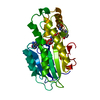
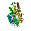
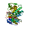
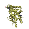
 Structure visualization
 Downloads & links
 Other downloads


 PDBj
PDBj Assembly

 Mass: 24.305 Da / Num. of mol.: 1 / Source method: obtained synthetically / Formula: Mg
 Mass: 18.015 Da / Num. of mol.: 366 / Source method: isolated from a natural source / Formula: H2O
 Sample preparation
 / Beamline: I04 / Wavelength: 0.8266
  Processing