[English] 日本語
Yorodumi
- PDB-2c32: Co-axial association of recombinant eye lens aquaporin-0 observed... -

+
Open data


ID or keywords:

Loading...

-
Basic information

Entry
Database: PDB / ID: 2c32
Title
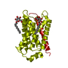
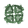
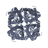
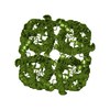
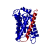
Co-axial association of recombinant eye lens aquaporin-0 observed in loosely packed 3D-crystals
ComponentsLENS FIBER MAJOR INTRINSIC PROTEIN
KeywordsMEMBRANE PROTEIN / EYE LENS / MIXED MICELLE / GAP JUNCTION / PHOSPHORYLATION / TRANSMEMBRANE
Function / homology
Function and homology information


Passive transport by Aquaporins / gap junction-mediated intercellular transport / water transport / water channel activity / structural constituent of eye lens / gap junction / lens development in camera-type eye / positive regulation of cell adhesion / visual perception / protein homotetramerization ...Passive transport by Aquaporins / gap junction-mediated intercellular transport / water transport / water channel activity / structural constituent of eye lens / gap junction / lens development in camera-type eye / positive regulation of cell adhesion / visual perception / protein homotetramerization / calmodulin binding / apical plasma membrane / endoplasmic reticulum / plasma membrane
Similarity search - Function
Aquaporin transporter / Major intrinsic protein, conserved site / MIP family signature. / Major intrinsic protein / Major intrinsic protein / Aquaporin-like
Similarity search - Domain/homology
Lens fiber major intrinsic protein
Similarity search - Component
Biological speciesBOS TAURUS (cattle)
MethodX-RAY DIFFRACTION / SYNCHROTRON / MOLECULAR REPLACEMENT / Resolution: 7.01 Å
AuthorsPalanivelu, D.V. / Schirmer, T.
CitationJournal: J.Mol.Biol. / Year: 2006
Title: Co-Axial Association of Recombinant Eye Lens Aquaporin-0 Observed in Loosely Packed 3D-Crystals
Authors: Palanivelu, D.V. / Kozono, D.E. / Engel, A. / Suda, K. / Lustig, A. / Agre, P. / Schirmer, T.
History
DepositionOct 3, 2005Deposition site: PDBE / Processing site: PDBE
Revision 1.0Dec 7, 2005Provider: repository / Type: Initial release
Revision 1.1May 7, 2011Group: Version format compliance
Revision 1.2Jul 13, 2011Group: Version format compliance
Revision 1.3Dec 13, 2023Group: Data collection / Database references ...Data collection / Database references / Other / Refinement description
Category: chem_comp_atom / chem_comp_bond ...chem_comp_atom / chem_comp_bond / database_2 / pdbx_database_status / pdbx_initial_refinement_model
Item: _database_2.pdbx_DOI / _database_2.pdbx_database_accession / _pdbx_database_status.status_code_sf

-
Structure visualization

Structure viewerMolecule:
MolmilJmol/JSmol

Downloads & links

-
Assembly

Deposited unit
A: LENS FIBER MAJOR INTRINSIC PROTEIN


Theoretical massNumber of molelcules
Total (without water)28,2451
Polymers28,2451
Non-polymers00
Water00
1
A: LENS FIBER MAJOR INTRINSIC PROTEIN

A: LENS FIBER MAJOR INTRINSIC PROTEIN

A: LENS FIBER MAJOR INTRINSIC PROTEIN

A: LENS FIBER MAJOR INTRINSIC PROTEIN


Theoretical massNumber of molelcules
Total (without water)112,9794
Polymers112,9794
Non-polymers00
Water0
TypeNameSymmetry operationNumber
identity operation1_555x,y,z1
crystal symmetry operation15_565y,-x+1,z1
crystal symmetry operation16_655-y+1,x,z1
crystal symmetry operation2_665-x+1,-y+1,z1
MethodPQS
Unit cell
Length a, b, c (Å)188.271, 188.271, 188.271
Angle α, β, γ (deg.)90.00, 90.00, 90.00
Int Tables number211
Space group name H-MI432

-
Components

#1: Protein LENS FIBER MAJOR INTRINSIC PROTEIN / AQUAPORIN-0 / AQP0 / MIP26 / MP26


Mass: 28244.865 Da / Num. of mol.: 1
Source method: isolated from a genetically manipulated source
Source: (gene. exp.) BOS TAURUS (cattle) / Organ: EYE / Plasmid: PYES2.0 / Production host: SACCHAROMYCES CEREVISIAE (brewer's yeast) / Strain (production host): SC334 / References: UniProt: P06624

-
Experimental details

-
Experiment

ExperimentMethod: X-RAY DIFFRACTION / Number of used crystals: 1

-
Sample preparation

CrystalDensity Matthews: 5.32 Å3/Da / Density % sol: 76.8 %
Crystal growpH: 8.9
Details: 100 MM BICINE NAOH PH 8.9, 350 MM NACL, 34 % (V/V) PEG400

-
Data collection

DiffractionMean temperature: 100 K
Diffraction sourceSource: SYNCHROTRON / Site: SLS / Beamline: X06SA / Wavelength: 0.9797
DetectorType: MARRESEARCH / Detector: IMAGE PLATE / Date: Aug 9, 2002
RadiationProtocol: SINGLE WAVELENGTH / Monochromatic (M) / Laue (L): M / Scattering type: x-ray
Radiation wavelengthWavelength: 0.9797 Å / Relative weight: 1
ReflectionResolution: 7.01→15 Å / Num. obs: 900 / % possible obs: 89.9 % / Observed criterion σ(I): 0 / Redundancy: 6.69 % / Rmerge(I) obs: 0.09 / Net I/σ(I): 5.21
Reflection shellResolution: 7.01→7.38 Å / Redundancy: 6.96 % / Rmerge(I) obs: 0.36 / Mean I/σ(I) obs: 2.06 / % possible all: 100

-
Processing

Software
NameVersionClassification
REFMAC5.2.0005refinement
MOSFLMdata reduction
SCALEPACKdata scaling
AMoREphasing
RefinementMethod to determine structure: MOLECULAR REPLACEMENT
Starting model: PDB ENTRY 1YMG
Resolution: 7.01→15 Å / Cor.coef. Fo:Fc: 0.756 / Cor.coef. Fo:Fc free: 0.781 / SU B: 517.315 / SU ML: 3.822 / Cross valid method: THROUGHOUT / σ(F): 0 / ESU R: 4.233 / ESU R Free: 4.204 / Stereochemistry target values: MAXIMUM LIKELIHOOD
Details: HYDROGENS HAVE BEEN ADDED IN THE RIDING POSITIONS. STRUCTURE SOLVED BY MOLECULAR REPLACEMENT AND RIGID BODY REFINEMENT (1 BODY, I.E. THE MONOMER OF THE CRYSTALLOGRAPHIC TETRAMER)
RfactorNum. reflection% reflectionSelection details
Rfree0.387 102 11.333 %RANDOM
Rwork0.39 ---
obs0.39 900 99.6 %-
Solvent computationIon probe radii: 0.8 Å / VDW probe radii: 1.2 Å / Solvent model: MASK BULK SOLVENT
Displacement parametersBiso mean: 51.75 Å2
Refinement stepCycle: LAST / Resolution: 7.01→15 Å
ProteinNucleic acidLigandSolventTotal
Num. atoms1761 0 0 0 1761
LS refinement shellResolution: 7.01→7.3 Å / Total num. of bins used: 10 /
RfactorNum. reflection
Rfree0.636 11
Rwork0.45 103

+
About Yorodumi

-
News

-
Feb 9, 2022. New format data for meta-information of EMDB entries

New format data for meta-information of EMDB entries

  • Version 3 of the EMDB header file is now the official format.
  • The previous official version 1.9 will be removed from the archive.

Related info.:EMDB header

External links:wwPDB to switch to version 3 of the EMDB data model

-
Aug 12, 2020. Covid-19 info

Covid-19 info

URL: https://pdbjlvh1.pdbj.org/emnavi/covid19.php

New page: Covid-19 featured information page in EM Navigator.

Related info.:Covid-19 info / Mar 5, 2020. Novel coronavirus structure data

+
Mar 5, 2020. Novel coronavirus structure data

Novel coronavirus structure data

Related info.:Yorodumi Speices / Aug 12, 2020. Covid-19 info

External links:COVID-19 featured content - PDBj / Molecule of the Month (242):Coronavirus Proteases

+
Jan 31, 2019. EMDB accession codes are about to change! (news from PDBe EMDB page)

EMDB accession codes are about to change! (news from PDBe EMDB page)

  • The allocation of 4 digits for EMDB accession codes will soon come to an end. Whilst these codes will remain in use, new EMDB accession codes will include an additional digit and will expand incrementally as the available range of codes is exhausted. The current 4-digit format prefixed with “EMD-” (i.e. EMD-XXXX) will advance to a 5-digit format (i.e. EMD-XXXXX), and so on. It is currently estimated that the 4-digit codes will be depleted around Spring 2019, at which point the 5-digit format will come into force.
  • The EM Navigator/Yorodumi systems omit the EMD- prefix.

Related info.:Q: What is EMD? / ID/Accession-code notation in Yorodumi/EM Navigator

External links:EMDB Accession Codes are Changing Soon! / Contact to PDBj

+
Jul 12, 2017. Major update of PDB

Major update of PDB

  • wwPDB released updated PDB data conforming to the new PDBx/mmCIF dictionary.
  • This is a major update changing the version number from 4 to 5, and with Remediation, in which all the entries are updated.
  • In this update, many items about electron microscopy experimental information are reorganized (e.g. em_software).
  • Now, EM Navigator and Yorodumi are based on the updated data.

External links:wwPDB Remediation / Enriched Model Files Conforming to OneDep Data Standards Now Available in the PDB FTP Archive

-
Yorodumi

Thousand views of thousand structures

  • Yorodumi is a browser for structure data from EMDB, PDB, SASBDB, etc.
  • This page is also the successor to EM Navigator detail page, and also detail information page/front-end page for Omokage search.
  • The word "yorodu" (or yorozu) is an old Japanese word meaning "ten thousand". "mi" (miru) is to see.

Related info.:EMDB / PDB / SASBDB / Comparison of 3 databanks / Yorodumi Search / Aug 31, 2016. New EM Navigator & Yorodumi / Yorodumi Papers / Jmol/JSmol / Function and homology information / Changes in new EM Navigator and Yorodumi

Read more