 Movie
Movie Controller

[English] 日本語
 Yorodumi
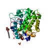
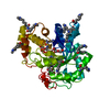
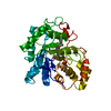
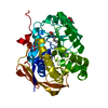
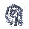
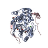
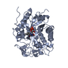
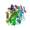
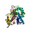
- PDB-4awt: Crystal structure of the reduced Shewanella Yellow Enzyme 1 (SYE1... -
+ Open data

- Basic information
| Entry | Database: PDB / ID: 4awt | ||||||
|---|---|---|---|---|---|---|---|
| Title | Crystal structure of the reduced Shewanella Yellow Enzyme 1 (SYE1) M25L mutant | ||||||
 Components Components | SYE1 | ||||||
 Keywords Keywords | OXIDOREDUCTASE / COFACTOR-BINDING | ||||||
| Function / homology |  Function and homology information Function and homology informationoxidoreductase activity, acting on the CH-CH group of donors, NAD or NADP as acceptor / FMN binding / cytosol Similarity search - Function | ||||||
| Biological species |  SHEWANELLA ONEIDENSIS (bacteria) SHEWANELLA ONEIDENSIS (bacteria) | ||||||
| Method |  X-RAY DIFFRACTION / SYNCHROTRON / MOLECULAR REPLACEMENT / Resolution: 0.98 Å X-RAY DIFFRACTION / SYNCHROTRON / MOLECULAR REPLACEMENT / Resolution: 0.98 Å | ||||||
 Authors Authors | Elegheert, J. / Brige, A. / Savvides, S.N. | ||||||
 Citation Citation | Journal: To be Published Title: Modulation of Isoalloxazine Ring Planarity Influences Fmn Electronic Properties in Old Yellow Enzymes Authors: Elegheert, J. / Pauwels, E. / Wille, G. / Brige, A. / Savvides, S.N. | ||||||
| History |
| ||||||
| Remark 700 | SHEET DETERMINATION METHOD: DSSP THE SHEETS PRESENTED AS "AB" IN EACH CHAIN ON SHEET RECORDS BELOW ... SHEET DETERMINATION METHOD: DSSP THE SHEETS PRESENTED AS "AB" IN EACH CHAIN ON SHEET RECORDS BELOW IS ACTUALLY AN 8-STRANDED BARREL THIS IS REPRESENTED BY A 9-STRANDED SHEET IN WHICH THE FIRST AND LAST STRANDS ARE IDENTICAL. |
- Structure visualization
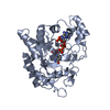
| Structure viewer | Molecule: MolmilJmol/JSmol |
|---|
- Downloads & links
-Download
| PDBx/mmCIF format | 4awt.cif.gz | 193.4 KB | Display | PDBx/mmCIF format |
|---|---|---|---|---|
| PDB format | pdb4awt.ent.gz | 151.6 KB | Display | PDB format |
| PDBx/mmJSON format | 4awt.json.gz | Tree view | PDBx/mmJSON format | |
| Others |  Other downloads Other downloads |
-Validation report
| Arichive directory | https://data.pdbj.org/pub/pdb/validation_reports/aw/4awtftp://data.pdbj.org/pub/pdb/validation_reports/aw/4awt | HTTPS FTP |
|---|
-Related structure data
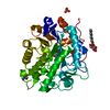
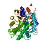
| Related structure data |  4awsSC 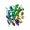 4awuC S: Starting model for refinement C: citing same article ( |
|---|---|
| Similar structure data |


-Links
 PDBj
PDBj- Assembly
| Deposited unit | 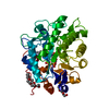
| ||||||||
|---|---|---|---|---|---|---|---|---|---|
| 1 |
| ||||||||
| Unit cell |
|
-Components
-Protein / Sugars , 2 types, 2 molecules A
| #1: Protein | Mass: 39699.629 Da / Num. of mol.: 1 / Mutation: YES Source method: isolated from a genetically manipulated source Details: N-TERMINAL T2 IS INVOLVED IN AN N, O FIVE RING ACETAL BY REACTION WITH HO-CH2-CH2-O-CH2-CHO, A PEG400 BREAKDOWN PRODUCT. Source: (gene. exp.) SHEWANELLA ONEIDENSIS (bacteria) / Strain: MR-1 / Plasmid: PACYC-DUET1 / Production host: |
|---|---|
| #4: Sugar | ChemComp-BOG /  Type: D-saccharide / Mass: 292.369 Da / Num. of mol.: 1 Type: D-saccharide / Mass: 292.369 Da / Num. of mol.: 1Source method: isolated from a genetically manipulated source Formula: C14H28O6 / Comment: detergent*YM |
-Non-polymers , 5 types, 625 molecules 


| #2: Chemical | ChemComp-FMN /  Mass: 456.344 Da / Num. of mol.: 1 / Source method: obtained synthetically / Formula: C17H21N4O9P Mass: 456.344 Da / Num. of mol.: 1 / Source method: obtained synthetically / Formula: C17H21N4O9P |
|---|---|
| #3: Chemical | ChemComp-SO4 /  Mass: 96.063 Da / Num. of mol.: 1 / Source method: obtained synthetically / Formula: SO4 Mass: 96.063 Da / Num. of mol.: 1 / Source method: obtained synthetically / Formula: SO4 |
| #5: Chemical | ChemComp-01F /  Mass: 90.078 Da / Num. of mol.: 1 / Source method: obtained synthetically / Formula: C3H6O3 Mass: 90.078 Da / Num. of mol.: 1 / Source method: obtained synthetically / Formula: C3H6O3 |
| #6: Chemical | ChemComp-PE4 /  Mass: 354.436 Da / Num. of mol.: 1 / Source method: obtained synthetically / Formula: C16H34O8 / Comment: precipitant*YM Mass: 354.436 Da / Num. of mol.: 1 / Source method: obtained synthetically / Formula: C16H34O8 / Comment: precipitant*YM |
| #7: Water | ChemComp-HOH / Mass: 18.015 Da / Num. of mol.: 621 / Source method: isolated from a natural source / Formula: H2O |
-Details
| Has protein modification | Y | ||
|---|---|---|---|
| Nonpolymer details | BETA-OCTYLGLUCO| Sequence details | M25L MUTATION TO MODULATE FMN PLANARITY | |
-Experimental details
-Experiment
| Experiment | Method: X-RAY DIFFRACTION / Number of used crystals: 1 |
|---|
- Sample preparation
| Crystal | Density Matthews: 2.25 Å3/Da / Density % sol: 45 % / Description: NONE |
|---|---|
| Crystal grow | pH: 8.2 Details: 100 MM TRIS PH 8.2, 1.65 M (NH4)2SO4, 2 % PEG400 AND 0.25 % (W/V) BETA-OCTYL GLUCOSIDE |
-Data collection
| Diffraction | Mean temperature: 100 K |
|---|---|
| Diffraction source | Source: SYNCHROTRON / Site: EMBL/DESY, HAMBURG  / Beamline: X13 / Wavelength: 0.8076 / Beamline: X13 / Wavelength: 0.8076 |
| Detector | Type: MARRESEARCH / Detector: CCD |
| Radiation | Protocol: SINGLE WAVELENGTH / Monochromatic (M) / Laue (L): M / Scattering type: x-ray |
| Radiation wavelength | Wavelength: 0.8076 Å / Relative weight: 1 |
| Reflection | Resolution: 0.98→30 Å / Num. obs: 202572 / % possible obs: 99 % / Redundancy: 4.5 % / Rmerge(I) obs: 0.05 / Net I/σ(I): 24.5 |
| Reflection shell | Resolution: 0.98→1 Å / Redundancy: 3.7 % / Rmerge(I) obs: 0.53 / Mean I/σ(I) obs: 3.9 / % possible all: 95.1 |
- Processing
| Software |
| |||||||||||||||||||||||||||||||||
|---|---|---|---|---|---|---|---|---|---|---|---|---|---|---|---|---|---|---|---|---|---|---|---|---|---|---|---|---|---|---|---|---|---|---|
| Refinement | Method to determine structure: MOLECULAR REPLACEMENT Starting model: PDB ENTRY 4AWS Resolution: 0.98→30 Å / Num. parameters: 33818 / Num. restraintsaints: 0 / Cross valid method: FREE R-VALUE / σ(F): 0 / Stereochemistry target values: ENGH AND HUBER
| |||||||||||||||||||||||||||||||||
| Refine analyze | Num. disordered residues: 46 / Occupancy sum hydrogen: 2802.34 / Occupancy sum non hydrogen: 3459 | |||||||||||||||||||||||||||||||||
| Refinement step | Cycle: LAST / Resolution: 0.98→30 Å
| |||||||||||||||||||||||||||||||||
| Refine LS restraints |
|
