 Movie
Movie Controller

[English] 日本語
 Yorodumi
- PDB-4awa: Crystal structure of active legumain in complex with YVAD-CMK at ... -
+ Open data

- Basic information
| Entry | Database: PDB / ID: 4awa | ||||||
|---|---|---|---|---|---|---|---|
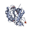
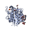
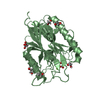
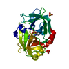
| Title | Crystal structure of active legumain in complex with YVAD-CMK at pH 5.0 | ||||||
 Components Components |
| ||||||
 Keywords Keywords | HYDROLASE/HYDROLASE INHIBITOR / HYDROLASE-HYDROLASE INHIBITOR COMPLEX / CYSTEINE PROTEASE / LYSOSOMAL / AEP / SUBSTRATE SPECIFICITY / MHCII / ANTIGEN PROCESSING / CANCER | ||||||
| Function / homology |  Function and homology information Function and homology informationnegative regulation of ERBB signaling pathway / legumain / vacuolar protein processing / antigen processing and presentation of exogenous peptide antigen via MHC class I, TAP-dependent / renal system process / Vitamin D (calciferol) metabolism / vitamin D metabolic process / receptor catabolic process / endolysosome lumen / Trafficking and processing of endosomal TLR ...negative regulation of ERBB signaling pathway / legumain / vacuolar protein processing / antigen processing and presentation of exogenous peptide antigen via MHC class I, TAP-dependent / renal system process / Vitamin D (calciferol) metabolism / vitamin D metabolic process / receptor catabolic process / endolysosome lumen / Trafficking and processing of endosomal TLR / positive regulation of monocyte chemotaxis / dendritic spine organization / positive regulation of endothelial cell chemotaxis / negative regulation of multicellular organism growth / response to acidic pH / cellular response to hepatocyte growth factor stimulus / associative learning / endopeptidase activator activity / MHC class II antigen presentation / positive regulation of mitotic cell cycle / lysosomal lumen / cellular response to calcium ion / : / positive regulation of long-term synaptic potentiation / protein maturation / antigen processing and presentation of exogenous peptide antigen via MHC class II / tau protein binding / cellular response to amyloid-beta / memory / apical part of cell / late endosome / peptidase activity / negative regulation of neuron apoptotic process / lysosome / negative regulation of gene expression / cysteine-type endopeptidase activity / positive regulation of cell population proliferation / perinuclear region of cytoplasm / proteolysis / extracellular exosome / extracellular region / cytoplasm Similarity search - Function | ||||||
| Biological species |  HOMO SAPIENS (human) HOMO SAPIENS (human)SYNTHETIC CONSTRUCT (others) | ||||||
| Method |  X-RAY DIFFRACTION / SYNCHROTRON / MOLECULAR REPLACEMENT / Resolution: 2.5 Å X-RAY DIFFRACTION / SYNCHROTRON / MOLECULAR REPLACEMENT / Resolution: 2.5 Å | ||||||
 Authors Authors | Dall, E. / Brandstetter, H. | ||||||
 Citation Citation | Journal: Proc.Natl.Acad.Sci.USA / Year: 2013 Title: Mechanistic and Structural Studies on Legumain Explain its Zymogenicity, Distinct Activation Pathways, and Regulation Authors: Dall, E. / Brandstetter, H. | ||||||
| History |
|
- Structure visualization
| Structure viewer | Molecule: MolmilJmol/JSmol |
|---|
- Downloads & links
-Download
| PDBx/mmCIF format | 4awa.cif.gz | 70.7 KB | Display | PDBx/mmCIF format |
|---|---|---|---|---|
| PDB format | pdb4awa.ent.gz | 51.5 KB | Display | PDB format |
| PDBx/mmJSON format | 4awa.json.gz | Tree view | PDBx/mmJSON format | |
| Others |  Other downloads Other downloads |
-Validation report
| Arichive directory | https://data.pdbj.org/pub/pdb/validation_reports/aw/4awaftp://data.pdbj.org/pub/pdb/validation_reports/aw/4awa | HTTPS FTP |
|---|
-Related structure data
| Related structure data | 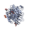 4aw9SC  4awbC 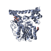 4fguC S: Starting model for refinement C: citing same article ( |
|---|---|
| Similar structure data |


-Links
 PDBj
PDBj


- Assembly
| Deposited unit | 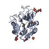
| |||||||||
|---|---|---|---|---|---|---|---|---|---|---|
| 1 |
| |||||||||
| Unit cell |
| |||||||||
| Components on special symmetry positions |
|
-Components
| #1: Protein | Mass: 32273.203 Da / Num. of mol.: 1 / Fragment: CATALYTIC DOMAIN, RESIDUES 26-309 / Mutation: YES Source method: isolated from a genetically manipulated source Source: (gene. exp.) HOMO SAPIENS (human) / Production host:  LEISHMANIA TARENTOLAE (eukaryote) / Strain (production host): P10 / References: UniProt: Q99538, legumain LEISHMANIA TARENTOLAE (eukaryote) / Strain (production host): P10 / References: UniProt: Q99538, legumain | ||||||||||
|---|---|---|---|---|---|---|---|---|---|---|---|
| #2: Protein/peptide | 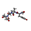  Type: Peptide-like / Class: Inhibitor / Mass: 524.996 Da / Num. of mol.: 1 / Source method: obtained synthetically / Source: (synth.) SYNTHETIC CONSTRUCT (others) / References: ACE-TYR-VAL-ALA-ASP-CHLOROMETHYLKETONE Type: Peptide-like / Class: Inhibitor / Mass: 524.996 Da / Num. of mol.: 1 / Source method: obtained synthetically / Source: (synth.) SYNTHETIC CONSTRUCT (others) / References: ACE-TYR-VAL-ALA-ASP-CHLOROMETHYLKETONE | ||||||||||
| #3: Chemical |   Mass: 96.063 Da / Num. of mol.: 2 / Source method: obtained synthetically / Formula: SO4 Mass: 96.063 Da / Num. of mol.: 2 / Source method: obtained synthetically / Formula: SO4#4: Sugar |   Type: D-saccharide, beta linking / Mass: 221.208 Da / Num. of mol.: 3 Type: D-saccharide, beta linking / Mass: 221.208 Da / Num. of mol.: 3Source method: isolated from a genetically manipulated source Formula: C8H15NO6 #5: Water | ChemComp-HOH / |  Mass: 18.015 Da / Num. of mol.: 112 / Source method: isolated from a natural source / Formula: H2O Mass: 18.015 Da / Num. of mol.: 112 / Source method: isolated from a natural source / Formula: H2OCompound details | ENGINEERED | Has protein modification | Y | Nonpolymer details | AC-YVAD-CMK (CHAIN I): AC-TYR-VAL-ALA-ASP-CHLOROMETH | |
-Experimental details
-Experiment
| Experiment | Method: X-RAY DIFFRACTION / Number of used crystals: 1 |
|---|
- Sample preparation
| Crystal | Density Matthews: 2.49 Å3/Da / Density % sol: 49 % / Description: NONE |
|---|---|
| Crystal grow | pH: 5 Details: 0.1 M CITRIC ACID PH 5.0, 0.2 M LITHIUM SULFATE MONOHYDRATE, 25% PEG 3350 |
-Data collection
| Diffraction | Mean temperature: 100 K |
|---|---|
| Diffraction source | Source: SYNCHROTRON / Site: ESRF  / Beamline: ID29 / Wavelength: 0.6999 / Beamline: ID29 / Wavelength: 0.6999 |
| Detector | Type: ADSC QUANTUM 315r / Detector: CCD |
| Radiation | Protocol: SINGLE WAVELENGTH / Monochromatic (M) / Laue (L): M / Scattering type: x-ray |
| Radiation wavelength | Wavelength: 0.6999 Å / Relative weight: 1 |
| Reflection | Resolution: 2.5→64.3 Å / Num. obs: 10650 / % possible obs: 94.9 % / Redundancy: 3.8 % / Rmerge(I) obs: 0.09 / Net I/σ(I): 16.5 |
| Reflection shell | Resolution: 2.5→7.91 Å / Redundancy: 3.7 % / Rmerge(I) obs: 0.23 / Mean I/σ(I) obs: 4 / % possible all: 95.2 |
- Processing
| Software |
| ||||||||||||||||||||||||||||||||||||||||||||||||||||||||||||||||||||||||||||||||||||||||||||||||||||||||||||||||||||||||||||||||||||||||||||||||||||||||||||||||||||||||||||||||||||||
|---|---|---|---|---|---|---|---|---|---|---|---|---|---|---|---|---|---|---|---|---|---|---|---|---|---|---|---|---|---|---|---|---|---|---|---|---|---|---|---|---|---|---|---|---|---|---|---|---|---|---|---|---|---|---|---|---|---|---|---|---|---|---|---|---|---|---|---|---|---|---|---|---|---|---|---|---|---|---|---|---|---|---|---|---|---|---|---|---|---|---|---|---|---|---|---|---|---|---|---|---|---|---|---|---|---|---|---|---|---|---|---|---|---|---|---|---|---|---|---|---|---|---|---|---|---|---|---|---|---|---|---|---|---|---|---|---|---|---|---|---|---|---|---|---|---|---|---|---|---|---|---|---|---|---|---|---|---|---|---|---|---|---|---|---|---|---|---|---|---|---|---|---|---|---|---|---|---|---|---|---|---|---|---|
| Refinement | Method to determine structure: MOLECULAR REPLACEMENT Starting model: PDB ENTRY 4AW9 Resolution: 2.5→64.3 Å / Cor.coef. Fo:Fc: 0.902 / Cor.coef. Fo:Fc free: 0.869 / SU B: 10.004 / SU ML: 0.23 / Cross valid method: THROUGHOUT / ESU R: 1.003 / ESU R Free: 0.316 / Stereochemistry target values: MAXIMUM LIKELIHOOD Details: HYDROGENS HAVE BEEN USED IF PRESENT IN THE INPUT. U VALUES REFINED INDIVIDUALLY
| ||||||||||||||||||||||||||||||||||||||||||||||||||||||||||||||||||||||||||||||||||||||||||||||||||||||||||||||||||||||||||||||||||||||||||||||||||||||||||||||||||||||||||||||||||||||
| Solvent computation | Ion probe radii: 0.8 Å / Shrinkage radii: 0.8 Å / VDW probe radii: 1.2 Å / Solvent model: MASK | ||||||||||||||||||||||||||||||||||||||||||||||||||||||||||||||||||||||||||||||||||||||||||||||||||||||||||||||||||||||||||||||||||||||||||||||||||||||||||||||||||||||||||||||||||||||
| Displacement parameters | Biso mean: 32.944 Å2
| ||||||||||||||||||||||||||||||||||||||||||||||||||||||||||||||||||||||||||||||||||||||||||||||||||||||||||||||||||||||||||||||||||||||||||||||||||||||||||||||||||||||||||||||||||||||
| Refinement step | Cycle: LAST / Resolution: 2.5→64.3 Å
| ||||||||||||||||||||||||||||||||||||||||||||||||||||||||||||||||||||||||||||||||||||||||||||||||||||||||||||||||||||||||||||||||||||||||||||||||||||||||||||||||||||||||||||||||||||||
| Refine LS restraints |
|