Mass: 39.098 Da / Num. of mol.: 2 / Source method: obtained synthetically / Formula: K
Compound details
ENGINEERED RESIDUE IN CHAIN A, VAL 353 TO LEU ENGINEERED RESIDUE IN CHAIN A, SER 395 TO GLY ...ENGINEERED RESIDUE IN CHAIN A, VAL 353 TO LEU ENGINEERED RESIDUE IN CHAIN A, SER 395 TO GLY ENGINEERED RESIDUE IN CHAIN B, VAL 353 TO LEU ENGINEERED RESIDUE IN CHAIN B, SER 395 TO GLY
Nonpolymer details
POTASSIUM ION (K): MODELLED INTO THE FO-FC PEAK AT 4 A. MAGNESIUM ION (MG): MODELLED INTO THE FO-FC ...POTASSIUM ION (K): MODELLED INTO THE FO-FC PEAK AT 4 A. MAGNESIUM ION (MG): MODELLED INTO THE FO-FC PEAK AROUND THE PO4 MOLECULES AT 4 A ACCORDING TO OTHER PPASE COMPLEXES.
-
Experimental details
-
Experiment
Experiment
Method: X-RAY DIFFRACTION / Number of used crystals: 1
-
Sample preparation
Crystal
Density Matthews: 3.24 Å3/Da / Density % sol: 62 % / Description: NONE
Crystal grow
Details: 35% PEG 400, 50 MM TRIS PH 8.0, 50 MM LISO4, 50 MM NASO4 PH range: PH 8.0
Monochromator: SI (111) MONOCHROMATOR / Protocol: SINGLE WAVELENGTH / Monochromatic (M) / Laue (L): M / Scattering type: x-ray
Radiation wavelength
Wavelength: 0.873 Å / Relative weight: 1
Reflection
Resolution: 4→30 Å / Num. obs: 17075 / % possible obs: 98.2 % / Observed criterion σ(I): -3 / Redundancy: 4.3 % / Biso Wilson estimate: 178.27 Å2 / Rmerge(I) obs: 0.09 / Net I/σ(I): 8.85
Reflection shell
Resolution: 4→4.22 Å / Redundancy: 3.5 % / Rmerge(I) obs: 0.97 / Mean I/σ(I) obs: 1.82 / % possible all: 96.8
-
Processing
Software
Name
Version
Classification
PHENIX
(PHENIX.REFINE)
refinement
XDS
datareduction
XDS
datascaling
PHASER
phasing
Refinement
Method to determine structure: MOLECULAR REPLACEMENT Starting model: HIGH RESOLUTION RESTING STATE TMPPASE STRUCTURE Resolution: 4→29.832 Å / SU ML: 1.7 / σ(F): 2.01 / Phase error: 43.81 / Stereochemistry target values: ML Details: SOME DISORDERED REGIONS AND SOME SIDE CHAINS WERE MODELLED BASED ON HIGH RESOLUTION RESTING TMPPASE STRUCTURE USED AS THE SEARCH MODEL.
Rfactor
Num. reflection
% reflection
Rfree
0.365
863
5.1 %
Rwork
0.2952
-
-
obs
0.2988
17061
98.88 %
Solvent computation
Shrinkage radii: 0.49 Å / VDW probe radii: 0.8 Å / Solvent model: FLAT BULK SOLVENT MODEL / Bsol: 154.483 Å2 / ksol: 0.235 e/Å3
Displacement parameters
Biso mean: 239 Å2
Baniso -1
Baniso -2
Baniso -3
1-
35.461 Å2
0 Å2
-23.7725 Å2
2-
-
-24.6168 Å2
0 Å2
3-
-
-
-10.8441 Å2
Refinement step
Cycle: LAST / Resolution: 4→29.832 Å
Protein
Nucleic acid
Ligand
Solvent
Total
Num. atoms
9376
0
30
0
9406
Refine LS restraints
Refine-ID
Type
Dev ideal
Number
X-RAY DIFFRACTION
f_bond_d
0.01
9606
X-RAY DIFFRACTION
f_angle_d
2.169
13080
X-RAY DIFFRACTION
f_dihedral_angle_d
16.051
3190
X-RAY DIFFRACTION
f_chiral_restr
0.111
1599
X-RAY DIFFRACTION
f_plane_restr
0.011
1611
Refine LS restraints NCS
Ens-ID
Dom-ID
Auth asym-ID
Number
Refine-ID
Type
Rms dev position (Å)
1
1
A
1833
X-RAY DIFFRACTION
POSITIONAL
1
2
B
1833
X-RAY DIFFRACTION
POSITIONAL
0.046
LS refinement shell
Resolution (Å)
Rfactor Rfree
Num. reflection Rfree
Rfactor Rwork
Num. reflection Rwork
Refine-ID
% reflection obs (%)
4-4.2499
0.3949
147
0.365
2659
X-RAY DIFFRACTION
98
4.2499-4.577
0.3795
136
0.314
2680
X-RAY DIFFRACTION
99
4.577-5.0356
0.4248
133
0.3006
2717
X-RAY DIFFRACTION
99
5.0356-5.7598
0.4344
157
0.3371
2682
X-RAY DIFFRACTION
99
5.7598-7.2395
0.3761
139
0.2833
2729
X-RAY DIFFRACTION
99
7.2395-29.8323
0.3237
151
0.2727
2731
X-RAY DIFFRACTION
99
Refinement TLS params.
Method: refined / Refine-ID: X-RAY DIFFRACTION
ID
L11 (°2)
L12 (°2)
L13 (°2)
L22 (°2)
L23 (°2)
L33 (°2)
S11 (Å °)
S12 (Å °)
S13 (Å °)
S21 (Å °)
S22 (Å °)
S23 (Å °)
S31 (Å °)
S32 (Å °)
S33 (Å °)
T11 (Å2)
T12 (Å2)
T13 (Å2)
T22 (Å2)
T23 (Å2)
T33 (Å2)
Origin x (Å)
Origin y (Å)
Origin z (Å)
1
0.393
0.061
0.1036
0.1886
-0.1319
0.269
0.2308
0.1653
-0.4616
-0.2723
0.211
-0.0114
0.3613
0.0411
0.6326
-0.7858
-0.3498
0.1615
0.458
0.1813
0.8781
9.3004
-0.2418
19.6878
2
0.5384
-0.3594
0.2117
0.7396
-0.1153
0.1414
-0.1429
-0.1305
0.2406
-0.0752
0.3207
-0.5918
-1.128
0.1023
0.0388
1.7622
-0.0936
-0.0371
0.6691
-0.0483
0.6781
4.1769
32.5716
39.5443
Refinement TLS group
ID
Refine-ID
Refine TLS-ID
Selection details
1
X-RAY DIFFRACTION
1
CHAINA
2
X-RAY DIFFRACTION
2
CHAINB
+
About Yorodumi
-
News
-
Feb 9, 2022. New format data for meta-information of EMDB entries
New format data for meta-information of EMDB entries
Version 3 of the EMDB header file is now the official format.
The previous official version 1.9 will be removed from the archive.
In the structure databanks used in Yorodumi, some data are registered as the other names, "COVID-19 virus" and "2019-nCoV". Here are the details of the virus and the list of structure data.
Jan 31, 2019. EMDB accession codes are about to change! (news from PDBe EMDB page)
EMDB accession codes are about to change! (news from PDBe EMDB page)
The allocation of 4 digits for EMDB accession codes will soon come to an end. Whilst these codes will remain in use, new EMDB accession codes will include an additional digit and will expand incrementally as the available range of codes is exhausted. The current 4-digit format prefixed with “EMD-” (i.e. EMD-XXXX) will advance to a 5-digit format (i.e. EMD-XXXXX), and so on. It is currently estimated that the 4-digit codes will be depleted around Spring 2019, at which point the 5-digit format will come into force.
The EM Navigator/Yorodumi systems omit the EMD- prefix.
Related info.:Q: What is EMD? / ID/Accession-code notation in Yorodumi/EM Navigator
Yorodumi is a browser for structure data from EMDB, PDB, SASBDB, etc.
This page is also the successor to EM Navigator detail page, and also detail information page/front-end page for Omokage search.
The word "yorodu" (or yorozu) is an old Japanese word meaning "ten thousand". "mi" (miru) is to see.
Related info.:EMDB / PDB / SASBDB / Comparison of 3 databanks / Yorodumi Search / Aug 31, 2016. New EM Navigator & Yorodumi / Yorodumi Papers / Jmol/JSmol / Function and homology information / Changes in new EM Navigator and Yorodumi
 Movie
Movie Controller

 Yorodumi
 Open data
 Basic information
 Components
 Keywords
 Function and homology information
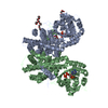
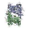
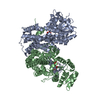
 THERMOTOGA MARITIMA (bacteria)
 X-RAY DIFFRACTION /
  Authors
 Citation
 Structure visualization
 Downloads & links
 Other downloads


 PDBj
PDBj Assembly


 Mass: 94.971 Da / Num. of mol.: 4 / Source method: obtained synthetically / Formula: PO4
 Mass: 24.305 Da / Num. of mol.: 8 / Source method: obtained synthetically / Formula: Mg
 Mass: 39.098 Da / Num. of mol.: 2 / Source method: obtained synthetically / Formula: K
 Sample preparation
 / Beamline: ID29 / Wavelength: 0.873
  Processing