 Movie
Movie Controller

[English] 日本語
 Yorodumi
- PDB-4amb: Crystal structure of the glycosyltransferase SnogD from Streptomy... -
+ Open data

- Basic information
| Entry | Database: PDB / ID: 4amb | ||||||
|---|---|---|---|---|---|---|---|
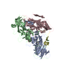
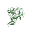
| Title | Crystal structure of the glycosyltransferase SnogD from Streptomyces nogalater | ||||||
 Components Components | SNOGD | ||||||
 Keywords Keywords | TRANSFERASE / POLYKETIDE BIOSYNTHESIS / GT1 FAMILY / NOGALAMYCIN | ||||||
| Function / homology |  Function and homology information Function and homology informationUDP-glycosyltransferase activity / hexosyltransferase activity / antibiotic biosynthetic process / identical protein binding Similarity search - Function | ||||||
| Biological species |  STREPTOMYCES NOGALATER (bacteria) STREPTOMYCES NOGALATER (bacteria) | ||||||
| Method |  X-RAY DIFFRACTION / SYNCHROTRON / MOLECULAR REPLACEMENT / Resolution: 2.62 Å X-RAY DIFFRACTION / SYNCHROTRON / MOLECULAR REPLACEMENT / Resolution: 2.62 Å | ||||||
 Authors Authors | Claesson, M. / Siitonen, V. / Dobritzsch, D. / Metsa-Ketela, M. / Schneider, G. | ||||||
 Citation Citation | Journal: FEBS J. / Year: 2012 Title: Crystal Structure of the Glycosyltransferase Snogd from the Biosynthetic Pathway of the Nogalamycin in Streptomyces Nogalater. Authors: Claesson, M. / Siitonen, V. / Dobritzsch, D. / Metsa-Ketela, M. / Schneider, G. | ||||||
| History |
|
- Structure visualization
| Structure viewer | Molecule: MolmilJmol/JSmol |
|---|
- Downloads & links
-Download
| PDBx/mmCIF format | 4amb.cif.gz | 148.5 KB | Display | PDBx/mmCIF format |
|---|---|---|---|---|
| PDB format | pdb4amb.ent.gz | 116.1 KB | Display | PDB format |
| PDBx/mmJSON format | 4amb.json.gz | Tree view | PDBx/mmJSON format | |
| Others |  Other downloads Other downloads |
-Validation report
| Arichive directory | https://data.pdbj.org/pub/pdb/validation_reports/am/4ambftp://data.pdbj.org/pub/pdb/validation_reports/am/4amb | HTTPS FTP |
|---|
-Related structure data
| Related structure data | 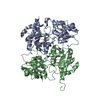 4amgC  4an4C 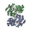 3d0qS S: Starting model for refinement C: citing same article ( |
|---|---|
| Similar structure data |
-Links
 PDBj
PDBj- Assembly
| Deposited unit | 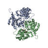
| ||||||||
|---|---|---|---|---|---|---|---|---|---|
| 1 |
| ||||||||
| Unit cell |
| ||||||||
| Noncrystallographic symmetry (NCS) | NCS oper: (Code: given Matrix: (-0.8528, -0.5222, -0.002406), Vector: |
-Components
| #1: Protein | Mass: 41946.566 Da / Num. of mol.: 2 / Fragment: RESIDUES 13-390 Source method: isolated from a genetically manipulated source Source: (gene. exp.) STREPTOMYCES NOGALATER (bacteria) / Plasmid: PNIC-BSAI / Production host: #2: Chemical | ChemComp-DUD / |   Mass: 388.162 Da / Num. of mol.: 1 / Source method: obtained synthetically / Formula: C9H14N2O11P2 Mass: 388.162 Da / Num. of mol.: 1 / Source method: obtained synthetically / Formula: C9H14N2O11P2#3: Water | ChemComp-HOH / |  Mass: 18.015 Da / Num. of mol.: 115 / Source method: isolated from a natural source / Formula: H2O Mass: 18.015 Da / Num. of mol.: 115 / Source method: isolated from a natural source / Formula: H2O |
|---|
-Experimental details
-Experiment
| Experiment | Method: X-RAY DIFFRACTION / Number of used crystals: 1 |
|---|
- Sample preparation
| Crystal | Density Matthews: 2.3 Å3/Da / Density % sol: 46.5 % / Description: NONE |
|---|---|
| Crystal grow | pH: 5.2 / Details: 26% (W/V) PEG 3350, 0.1 M CITRATE PH 5.2 |
-Data collection
| Diffraction | Mean temperature: 100 K |
|---|---|
| Diffraction source | Source: SYNCHROTRON / Site: ESRF  / Beamline: ID14-1 / Wavelength: 0.934 / Beamline: ID14-1 / Wavelength: 0.934 |
| Detector | Type: ADSC QUANTUM 315r / Detector: CCD |
| Radiation | Protocol: SINGLE WAVELENGTH / Monochromatic (M) / Laue (L): M / Scattering type: x-ray |
| Radiation wavelength | Wavelength: 0.934 Å / Relative weight: 1 |
| Reflection | Resolution: 2.62→60 Å / Num. obs: 23310 / % possible obs: 97.6 % / Observed criterion σ(I): 2 / Redundancy: 3 % / Rmerge(I) obs: 0.14 / Net I/σ(I): 7.9 |
| Reflection shell | Resolution: 2.62→2.76 Å / Redundancy: 2.3 % / Rmerge(I) obs: 0.46 / Mean I/σ(I) obs: 2 / % possible all: 86.7 |
- Processing
| Software |
| ||||||||||||||||||||||||||||||||||||||||||||||||||||||||||||||||||||||||||||||||||||||||||||||||||||||||||||||||||||||||||||||||||||||||||||||||||||||||||||||||||||||||||||||||||||||
|---|---|---|---|---|---|---|---|---|---|---|---|---|---|---|---|---|---|---|---|---|---|---|---|---|---|---|---|---|---|---|---|---|---|---|---|---|---|---|---|---|---|---|---|---|---|---|---|---|---|---|---|---|---|---|---|---|---|---|---|---|---|---|---|---|---|---|---|---|---|---|---|---|---|---|---|---|---|---|---|---|---|---|---|---|---|---|---|---|---|---|---|---|---|---|---|---|---|---|---|---|---|---|---|---|---|---|---|---|---|---|---|---|---|---|---|---|---|---|---|---|---|---|---|---|---|---|---|---|---|---|---|---|---|---|---|---|---|---|---|---|---|---|---|---|---|---|---|---|---|---|---|---|---|---|---|---|---|---|---|---|---|---|---|---|---|---|---|---|---|---|---|---|---|---|---|---|---|---|---|---|---|---|---|
| Refinement | Method to determine structure: MOLECULAR REPLACEMENT Starting model: PDB ENTRY 3D0Q Resolution: 2.62→50 Å / Cor.coef. Fo:Fc: 0.908 / Cor.coef. Fo:Fc free: 0.83 / SU B: 14.565 / SU ML: 0.307 / Cross valid method: THROUGHOUT / ESU R Free: 0.402 / Stereochemistry target values: MAXIMUM LIKELIHOOD Details: HYDROGENS HAVE BEEN ADDED IN THE RIDING POSITIONS. U VALUES REFINED INDIVIDUALLY
| ||||||||||||||||||||||||||||||||||||||||||||||||||||||||||||||||||||||||||||||||||||||||||||||||||||||||||||||||||||||||||||||||||||||||||||||||||||||||||||||||||||||||||||||||||||||
| Solvent computation | Ion probe radii: 0.8 Å / Shrinkage radii: 0.8 Å / VDW probe radii: 1.2 Å / Solvent model: MASK | ||||||||||||||||||||||||||||||||||||||||||||||||||||||||||||||||||||||||||||||||||||||||||||||||||||||||||||||||||||||||||||||||||||||||||||||||||||||||||||||||||||||||||||||||||||||
| Displacement parameters | Biso mean: 27.945 Å2
| ||||||||||||||||||||||||||||||||||||||||||||||||||||||||||||||||||||||||||||||||||||||||||||||||||||||||||||||||||||||||||||||||||||||||||||||||||||||||||||||||||||||||||||||||||||||
| Refinement step | Cycle: LAST / Resolution: 2.62→50 Å
| ||||||||||||||||||||||||||||||||||||||||||||||||||||||||||||||||||||||||||||||||||||||||||||||||||||||||||||||||||||||||||||||||||||||||||||||||||||||||||||||||||||||||||||||||||||||
| Refine LS restraints |
|