 Movie
Movie Controller

[English] 日本語
 Yorodumi
- PDB-4ahv: Parallel screening of a low molecular weight compound library: do... -
+ Open data

- Basic information
| Entry | Database: PDB / ID: 4ahv | ||||||
|---|---|---|---|---|---|---|---|
| Title | Parallel screening of a low molecular weight compound library: do differences in methodology affect hit identification | ||||||
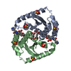
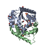
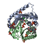
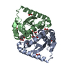
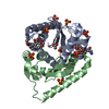
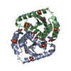
 Components Components | INTEGRASE | ||||||
 Keywords Keywords | TRANSFERASE | ||||||
| Function / homology |  Function and homology information Function and homology informationHIV-1 retropepsin / symbiont-mediated activation of host apoptosis / retroviral ribonuclease H / exoribonuclease H / exoribonuclease H activity / DNA integration / viral genome integration into host DNA / establishment of integrated proviral latency / RNA-directed DNA polymerase / RNA stem-loop binding ...HIV-1 retropepsin / symbiont-mediated activation of host apoptosis / retroviral ribonuclease H / exoribonuclease H / exoribonuclease H activity / DNA integration / viral genome integration into host DNA / establishment of integrated proviral latency / RNA-directed DNA polymerase / RNA stem-loop binding / viral penetration into host nucleus / host multivesicular body / RNA-directed DNA polymerase activity / RNA-DNA hybrid ribonuclease activity / Transferases; Transferring phosphorus-containing groups; Nucleotidyltransferases / host cell / viral nucleocapsid / DNA recombination / DNA-directed DNA polymerase / aspartic-type endopeptidase activity / Hydrolases; Acting on ester bonds / DNA-directed DNA polymerase activity / symbiont-mediated suppression of host gene expression / viral translational frameshifting / symbiont entry into host cell / lipid binding / host cell nucleus / host cell plasma membrane / virion membrane / structural molecule activity / proteolysis / DNA binding / zinc ion binding Similarity search - Function | ||||||
| Biological species |   HUMAN IMMUNODEFICIENCY VIRUS HUMAN IMMUNODEFICIENCY VIRUS | ||||||
| Method |  X-RAY DIFFRACTION / SYNCHROTRON / MOLECULAR REPLACEMENT / Resolution: 1.8 Å X-RAY DIFFRACTION / SYNCHROTRON / MOLECULAR REPLACEMENT / Resolution: 1.8 Å | ||||||
 Authors Authors | Wielens, J. / Heady, S.J. / Rhodes, D.I. / Mulder, R.J. / Dolezal, O. / Deadman, J.J. / Newman, J. / Chalmers, D.K. / Parker, M.W. / Peat, T.S. / Scanlon, M.J. | ||||||
 Citation Citation | Journal: J.Biomol.Screen / Year: 2013 Title: Parallel Screening of Low Molecular Weight Fragment Libraries: Do Differences in Methodology Affect Hit Identification? Authors: Wielens, J. / Headey, S.J. / Rhodes, D.I. / Mulder, R.J. / Dolezal, O. / Deadman, J.J. / Newman, J. / Chalmers, D.K. / Parker, M.W. / Peat, T.S. / Scanlon, M.J. | ||||||
| History |
|
- Structure visualization
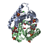
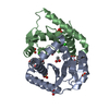
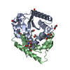
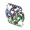
| Structure viewer | Molecule: MolmilJmol/JSmol |
|---|
- Downloads & links
-Download
| PDBx/mmCIF format | 4ahv.cif.gz | 83.8 KB | Display | PDBx/mmCIF format |
|---|---|---|---|---|
| PDB format | pdb4ahv.ent.gz | 62.5 KB | Display | PDB format |
| PDBx/mmJSON format | 4ahv.json.gz | Tree view | PDBx/mmJSON format | |
| Others |  Other downloads Other downloads |
-Validation report
| Arichive directory | https://data.pdbj.org/pub/pdb/validation_reports/ah/4ahvftp://data.pdbj.org/pub/pdb/validation_reports/ah/4ahv | HTTPS FTP |
|---|
-Related structure data
| Related structure data | 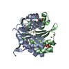 3vq4C 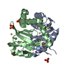 3vq5C 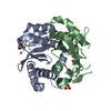 3vq6C 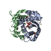 3vq7C  3vq8C 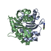 3vq9C  3vqaC 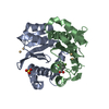 3vqbC 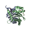 3vqcC 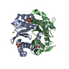 3vqdC 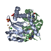 3vqeC 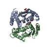 3vqpC 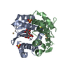 3vqqC 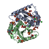 4ah9C 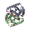 4ahrSC 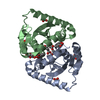 4ahsC 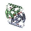 4ahtC 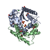 4ahuC C: citing same article ( S: Starting model for refinement |
|---|---|
| Similar structure data |
-Links
 PDBj
PDBj


- Assembly
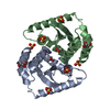
| Deposited unit | 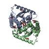
| ||||||||
|---|---|---|---|---|---|---|---|---|---|
| 1 |
| ||||||||
| Unit cell |
|
-Components
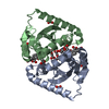
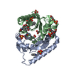
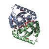
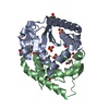
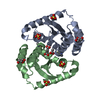
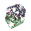
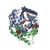
-Protein , 1 types, 2 molecules AB
| #1: Protein | Mass: 20044.672 Da / Num. of mol.: 2 / Fragment: CATALYTIC DOMAIN, RESIDUES 1197-1359 / Mutation: YES Source method: isolated from a genetically manipulated source Source: (gene. exp.) HUMAN IMMUNODEFICIENCY VIRUS / Strain: TYPE 1 / Production host:  References: UniProt: P12497, nicotinamide-nucleotide adenylyltransferase |
|---|
-Non-polymers , 6 types, 188 molecules 


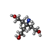
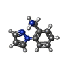

| #2: Chemical | ChemComp-SO4 /  Mass: 96.063 Da / Num. of mol.: 6 / Source method: obtained synthetically / Formula: SO4 Mass: 96.063 Da / Num. of mol.: 6 / Source method: obtained synthetically / Formula: SO4#3: Chemical |  Mass: 60.052 Da / Num. of mol.: 2 / Source method: obtained synthetically / Formula: C2H4O2 Mass: 60.052 Da / Num. of mol.: 2 / Source method: obtained synthetically / Formula: C2H4O2#4: Chemical | ChemComp-EDO /  Mass: 62.068 Da / Num. of mol.: 5 / Source method: obtained synthetically / Formula: C2H6O2 Mass: 62.068 Da / Num. of mol.: 5 / Source method: obtained synthetically / Formula: C2H6O2#5: Chemical |  Mass: 163.215 Da / Num. of mol.: 2 / Source method: obtained synthetically / Formula: C7H17NO3 / Comment: pH buffer*YM Mass: 163.215 Da / Num. of mol.: 2 / Source method: obtained synthetically / Formula: C7H17NO3 / Comment: pH buffer*YM#6: Chemical |  Mass: 173.214 Da / Num. of mol.: 2 / Source method: obtained synthetically / Formula: C10H11N3 Mass: 173.214 Da / Num. of mol.: 2 / Source method: obtained synthetically / Formula: C10H11N3#7: Water | ChemComp-HOH / | Mass: 18.015 Da / Num. of mol.: 171 / Source method: isolated from a natural source / Formula: H2O |
|---|
-Details
| Compound details | ENGINEERED RESIDUE IN CHAIN A, CYS1203 TO SER ENGINEERED RESIDUE IN CHAIN A, PHE1286 TO ASP ...ENGINEERED |
|---|
-Experimental details
-Experiment
| Experiment | Method: X-RAY DIFFRACTION / Number of used crystals: 20 |
|---|
- Sample preparation
| Crystal | Density Matthews: 2.94 Å3/Da / Density % sol: 58.2 % / Description: NONE |
|---|---|
| Crystal grow | Temperature: 293 K / Method: vapor diffusion, sitting drop / pH: 5.5 Details: PROTEIN: 5.5MG/ML IN 40 MM TRIS BUFFER AT PH 8.0, 250 MM NACL, 30 MM MGCL2, 5MM DTT. CRYSTALLANT: 100 MM SODIUM ACETATE PH 5.0 TO 5.5, 1.2 TO 1.5 M AMMONIUM SULFATE AT 20C IN SITTING DROP ...Details: PROTEIN: 5.5MG/ML IN 40 MM TRIS BUFFER AT PH 8.0, 250 MM NACL, 30 MM MGCL2, 5MM DTT. CRYSTALLANT: 100 MM SODIUM ACETATE PH 5.0 TO 5.5, 1.2 TO 1.5 M AMMONIUM SULFATE AT 20C IN SITTING DROP PLATES. FRAGMENTS WERE SOAKED INTO PREFORMED CRYSTALS 24-48 HOURS PRIOR TO DATA COLLECTION. |
-Data collection
| Diffraction | Mean temperature: 100 K |
|---|---|
| Diffraction source | Source: SYNCHROTRON / Site: Australian Synchrotron  / Beamline: MX1 / Wavelength: 0.9531 / Beamline: MX1 / Wavelength: 0.9531 |
| Detector | Type: ADSC QUANTUM 210r / Detector: CCD / Date: Apr 25, 2008 |
| Radiation | Protocol: SINGLE WAVELENGTH / Monochromatic (M) / Laue (L): M / Scattering type: x-ray |
| Radiation wavelength | Wavelength: 0.9531 Å / Relative weight: 1 |
| Reflection | Resolution: 1.8→61.4 Å / Num. obs: 33604 / % possible obs: 96.6 % / Observed criterion σ(I): 1 / Redundancy: 3.9 % / Rmerge(I) obs: 0.08 / Net I/σ(I): 12.3 |
| Reflection shell | Resolution: 1.8→1.9 Å / Redundancy: 2.4 % / Rmerge(I) obs: 0.82 / Mean I/σ(I) obs: 1.1 / % possible all: 81.3 |
- Processing
| Software |
| ||||||||||||||||||||||||||||||||||||||||||||||||||||||||||||||||||||||||||||||||||||||||||||||||||||||||||||||||||||||||||||||||||||||||||||||||||||||||||||||||||||||||||||||||||||||
|---|---|---|---|---|---|---|---|---|---|---|---|---|---|---|---|---|---|---|---|---|---|---|---|---|---|---|---|---|---|---|---|---|---|---|---|---|---|---|---|---|---|---|---|---|---|---|---|---|---|---|---|---|---|---|---|---|---|---|---|---|---|---|---|---|---|---|---|---|---|---|---|---|---|---|---|---|---|---|---|---|---|---|---|---|---|---|---|---|---|---|---|---|---|---|---|---|---|---|---|---|---|---|---|---|---|---|---|---|---|---|---|---|---|---|---|---|---|---|---|---|---|---|---|---|---|---|---|---|---|---|---|---|---|---|---|---|---|---|---|---|---|---|---|---|---|---|---|---|---|---|---|---|---|---|---|---|---|---|---|---|---|---|---|---|---|---|---|---|---|---|---|---|---|---|---|---|---|---|---|---|---|---|---|
| Refinement | Method to determine structure: MOLECULAR REPLACEMENT Starting model: PDB ENTRY 4AHR Resolution: 1.8→61.43 Å / Cor.coef. Fo:Fc: 0.961 / Cor.coef. Fo:Fc free: 0.932 / SU B: 2.613 / SU ML: 0.08 / Cross valid method: THROUGHOUT / ESU R: 0.117 / ESU R Free: 0.122 / Stereochemistry target values: MAXIMUM LIKELIHOOD Details: HYDROGENS HAVE BEEN ADDED IN THE RIDING POSITIONS. HYDROGENS HAVE BEEN USED IF PRESENT IN THE INPUT. U VALUES REFINED INDIVIDUALLY
| ||||||||||||||||||||||||||||||||||||||||||||||||||||||||||||||||||||||||||||||||||||||||||||||||||||||||||||||||||||||||||||||||||||||||||||||||||||||||||||||||||||||||||||||||||||||
| Solvent computation | Ion probe radii: 0.8 Å / Shrinkage radii: 0.8 Å / VDW probe radii: 1.2 Å / Solvent model: MASK | ||||||||||||||||||||||||||||||||||||||||||||||||||||||||||||||||||||||||||||||||||||||||||||||||||||||||||||||||||||||||||||||||||||||||||||||||||||||||||||||||||||||||||||||||||||||
| Displacement parameters | Biso mean: 26.12 Å2
| ||||||||||||||||||||||||||||||||||||||||||||||||||||||||||||||||||||||||||||||||||||||||||||||||||||||||||||||||||||||||||||||||||||||||||||||||||||||||||||||||||||||||||||||||||||||
| Refinement step | Cycle: LAST / Resolution: 1.8→61.43 Å
| ||||||||||||||||||||||||||||||||||||||||||||||||||||||||||||||||||||||||||||||||||||||||||||||||||||||||||||||||||||||||||||||||||||||||||||||||||||||||||||||||||||||||||||||||||||||
| Refine LS restraints |
|