 Movie
Movie Controller

[English] 日本語
 Yorodumi
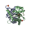
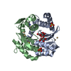
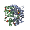
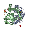
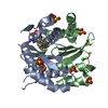
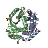
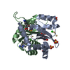
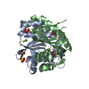
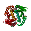
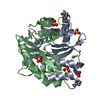
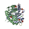
- PDB-3vq8: HIV-1 IN core domain in complex with (3R)-3,4-dihydro-2H-chromen-... -
+ Open data

- Basic information
| Entry | Database: PDB / ID: 3vq8 | ||||||
|---|---|---|---|---|---|---|---|
| Title | HIV-1 IN core domain in complex with (3R)-3,4-dihydro-2H-chromen-3-ylmethanol | ||||||
 Components Components | POL polyprotein | ||||||
 Keywords Keywords | TRANSFERASE/TRANSFERASE INHIBITOR / RNaseH / DNA binding / DNA cleavage / DNA integration / TRANSFERASE-TRANSFERASE INHIBITOR complex | ||||||
| Function / homology |  Function and homology information Function and homology informationexoribonuclease H activity / DNA integration / viral genome integration into host DNA / establishment of integrated proviral latency / RNA stem-loop binding / viral penetration into host nucleus / host multivesicular body / RNA-directed DNA polymerase activity / RNA-DNA hybrid ribonuclease activity / host cell ...exoribonuclease H activity / DNA integration / viral genome integration into host DNA / establishment of integrated proviral latency / RNA stem-loop binding / viral penetration into host nucleus / host multivesicular body / RNA-directed DNA polymerase activity / RNA-DNA hybrid ribonuclease activity / host cell / viral nucleocapsid / DNA recombination / aspartic-type endopeptidase activity / DNA-directed DNA polymerase activity / symbiont-mediated suppression of host gene expression / viral translational frameshifting / symbiont entry into host cell / lipid binding / host cell nucleus / host cell plasma membrane / virion membrane / proteolysis / DNA binding / zinc ion binding Similarity search - Function | ||||||
| Biological species |   Human immunodeficiency virus 1 Human immunodeficiency virus 1 | ||||||
| Method |  X-RAY DIFFRACTION / SYNCHROTRON / MOLECULAR REPLACEMENT / Resolution: 1.6 Å X-RAY DIFFRACTION / SYNCHROTRON / MOLECULAR REPLACEMENT / Resolution: 1.6 Å | ||||||
 Authors Authors | Wielens, J. / Chalmers, D.K. / Parker, M.W. / Scanlon, M.J. | ||||||
 Citation Citation | Journal: J Biomol Screen / Year: 2013 Title: Parallel screening of low molecular weight fragment libraries: do differences in methodology affect hit identification? Authors: Wielens, J. / Headey, S.J. / Rhodes, D.I. / Mulder, R.J. / Dolezal, O. / Deadman, J.J. / Newman, J. / Chalmers, D.K. / Parker, M.W. / Peat, T.S. / Scanlon, M.J. | ||||||
| History |
|
- Structure visualization
| Structure viewer | Molecule: MolmilJmol/JSmol |
|---|
- Downloads & links
-Download
| PDBx/mmCIF format | 3vq8.cif.gz | 70.5 KB | Display | PDBx/mmCIF format |
|---|---|---|---|---|
| PDB format | pdb3vq8.ent.gz | 50.5 KB | Display | PDB format |
| PDBx/mmJSON format | 3vq8.json.gz | Tree view | PDBx/mmJSON format | |
| Others |  Other downloads Other downloads |
-Validation report
| Arichive directory | https://data.pdbj.org/pub/pdb/validation_reports/vq/3vq8ftp://data.pdbj.org/pub/pdb/validation_reports/vq/3vq8 | HTTPS FTP |
|---|
-Related structure data
| Related structure data | 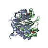 3vq4C  3vq5C 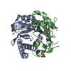 3vq6C 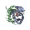 3vq7C 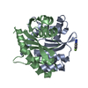 3vq9C  3vqaC 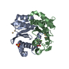 3vqbC 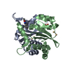 3vqcC 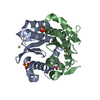 3vqdC 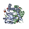 3vqeC 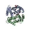 3vqpC 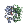 3vqqC 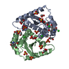 4ah9C 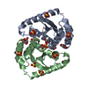 4ahrC 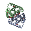 4ahsC 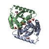 4ahtC 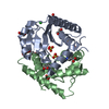 4ahuC 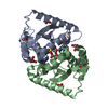 4ahvC 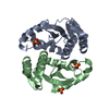 3l3uS S: Starting model for refinement C: citing same article ( |
|---|---|
| Similar structure data |

-Links
 PDBj
PDBj- Assembly
| Deposited unit | 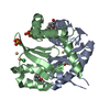
| ||||||||
|---|---|---|---|---|---|---|---|---|---|
| 1 |
| ||||||||
| Unit cell |
|
-Components
-Protein , 1 types, 2 molecules AB
| #1: Protein | Mass: 17101.393 Da / Num. of mol.: 2 / Fragment: integrase core domain, UNP residues 771-927 / Mutation: C56S, S123G, T124A, K127R, W131D, F139D, F185H Source method: isolated from a genetically manipulated source Source: (gene. exp.) Human immunodeficiency virus 1 / Strain: NL43 / Gene: pol / Plasmid: PeT23b+ / Production host:  |
|---|
-Non-polymers , 5 types, 80 molecules 


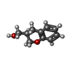

| #2: Chemical | ChemComp-CD /  Mass: 112.411 Da / Num. of mol.: 4 / Source method: obtained synthetically / Formula: Cd Mass: 112.411 Da / Num. of mol.: 4 / Source method: obtained synthetically / Formula: Cd#3: Chemical |  Mass: 35.453 Da / Num. of mol.: 2 / Source method: obtained synthetically / Formula: Cl Mass: 35.453 Da / Num. of mol.: 2 / Source method: obtained synthetically / Formula: Cl#4: Chemical | ChemComp-SO4 /  Mass: 96.063 Da / Num. of mol.: 4 / Source method: obtained synthetically / Formula: SO4 Mass: 96.063 Da / Num. of mol.: 4 / Source method: obtained synthetically / Formula: SO4#5: Chemical |  Mass: 164.201 Da / Num. of mol.: 2 / Source method: obtained synthetically / Formula: C10H12O2 Mass: 164.201 Da / Num. of mol.: 2 / Source method: obtained synthetically / Formula: C10H12O2#6: Water | ChemComp-HOH / | Mass: 18.015 Da / Num. of mol.: 68 / Source method: isolated from a natural source / Formula: H2O |
|---|
-Experimental details
-Experiment
| Experiment | Method: X-RAY DIFFRACTION / Number of used crystals: 1 |
|---|
- Sample preparation
| Crystal | Density Matthews: 2.11 Å3/Da / Density % sol: 41.63 % |
|---|---|
| Crystal grow | Temperature: 295 K / Method: vapor diffusion, hanging drop / pH: 4.6 Details: 1.6M AmSO4, 0.1M Na Citrate pH 4.6, 50mM CdCl2, VAPOR DIFFUSION, HANGING DROP, temperature 295K |
-Data collection
| Diffraction | Mean temperature: 100 K |
|---|---|
| Diffraction source | Source: SYNCHROTRON / Site: Australian Synchrotron  / Beamline: MX1 / Wavelength: 0.9544 Å / Beamline: MX1 / Wavelength: 0.9544 Å |
| Detector | Type: ADSC QUANTUM 210r / Detector: CCD / Date: May 15, 2009 / Details: mirror |
| Radiation | Monochromator: double mirror / Protocol: SINGLE WAVELENGTH / Monochromatic (M) / Laue (L): M / Scattering type: x-ray |
| Radiation wavelength | Wavelength: 0.9544 Å / Relative weight: 1 |
| Reflection | Resolution: 1.44→50 Å / Num. all: 50498 / Num. obs: 47885 / % possible obs: 94.8 % / Observed criterion σ(F): 2 / Observed criterion σ(I): 2 / Redundancy: 5.2 % / Rmerge(I) obs: 0.119 / Rsym value: 0.081 / Net I/σ(I): 12.74 |
| Reflection shell | Resolution: 1.44→1.53 Å / Redundancy: 3.9 % / Rmerge(I) obs: 0.793 / Mean I/σ(I) obs: 2.4 / Num. unique all: 7027 / Rsym value: 0.706 / % possible all: 85.9 |
- Processing
| Software |
| ||||||||||||||||||||||||||||||||||||||||||||||||||||||||||||||||||||||||||||||||||||||||||||||||||||||||||||||||||||||||||||||||||||||||||||||||||||||||||||||||||||||||||
|---|---|---|---|---|---|---|---|---|---|---|---|---|---|---|---|---|---|---|---|---|---|---|---|---|---|---|---|---|---|---|---|---|---|---|---|---|---|---|---|---|---|---|---|---|---|---|---|---|---|---|---|---|---|---|---|---|---|---|---|---|---|---|---|---|---|---|---|---|---|---|---|---|---|---|---|---|---|---|---|---|---|---|---|---|---|---|---|---|---|---|---|---|---|---|---|---|---|---|---|---|---|---|---|---|---|---|---|---|---|---|---|---|---|---|---|---|---|---|---|---|---|---|---|---|---|---|---|---|---|---|---|---|---|---|---|---|---|---|---|---|---|---|---|---|---|---|---|---|---|---|---|---|---|---|---|---|---|---|---|---|---|---|---|---|---|---|---|---|---|---|---|
| Refinement | Method to determine structure: MOLECULAR REPLACEMENT Starting model: PDB ENTRY 3l3u Resolution: 1.6→26.8 Å / Cor.coef. Fo:Fc: 0.93 / Cor.coef. Fo:Fc free: 0.921 / SU B: 1.877 / SU ML: 0.069 / Cross valid method: THROUGHOUT / ESU R: 0.114 / ESU R Free: 0.11 / Stereochemistry target values: MAXIMUM LIKELIHOOD / Details: HYDROGENS HAVE BEEN USED IF PRESENT IN THE INPUT
| ||||||||||||||||||||||||||||||||||||||||||||||||||||||||||||||||||||||||||||||||||||||||||||||||||||||||||||||||||||||||||||||||||||||||||||||||||||||||||||||||||||||||||
| Solvent computation | Ion probe radii: 0.8 Å / Shrinkage radii: 0.8 Å / VDW probe radii: 1.2 Å / Solvent model: MASK | ||||||||||||||||||||||||||||||||||||||||||||||||||||||||||||||||||||||||||||||||||||||||||||||||||||||||||||||||||||||||||||||||||||||||||||||||||||||||||||||||||||||||||
| Displacement parameters | Biso mean: 22.344 Å2
| ||||||||||||||||||||||||||||||||||||||||||||||||||||||||||||||||||||||||||||||||||||||||||||||||||||||||||||||||||||||||||||||||||||||||||||||||||||||||||||||||||||||||||
| Refinement step | Cycle: LAST / Resolution: 1.6→26.8 Å
| ||||||||||||||||||||||||||||||||||||||||||||||||||||||||||||||||||||||||||||||||||||||||||||||||||||||||||||||||||||||||||||||||||||||||||||||||||||||||||||||||||||||||||
| Refine LS restraints |
| ||||||||||||||||||||||||||||||||||||||||||||||||||||||||||||||||||||||||||||||||||||||||||||||||||||||||||||||||||||||||||||||||||||||||||||||||||||||||||||||||||||||||||
| LS refinement shell | Resolution: 1.6→1.642 Å / Total num. of bins used: 20
|
