 Movie
Movie Controller


+ Open data

- Basic information
| Entry | Database: PDB / ID: 4aey | ||||||
|---|---|---|---|---|---|---|---|
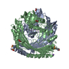
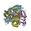
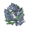
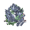
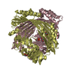
| Title | Crystal structure of FolX from Pseudomonas aeruginosa | ||||||
 Components Components | D-ERYTHRO-7,8-DIHYDRONEOPTERIN TRIPHOSPHATE EPIMERASE | ||||||
 Keywords Keywords | ISOMERASE / PTERIDINE BIOSYNTHESIS | ||||||
| Function / homology |  Function and homology information Function and homology informationdihydroneopterin triphosphate 2'-epimerase / dihydroneopterin triphosphate 2'-epimerase activity / dihydroneopterin aldolase activity / folic acid-containing compound metabolic process / tetrahydrobiopterin biosynthetic process / cytosol / cytoplasm Similarity search - Function | ||||||
| Biological species |   PSEUDOMONAS AERUGINOSA (bacteria) PSEUDOMONAS AERUGINOSA (bacteria) | ||||||
| Method |  X-RAY DIFFRACTION / SYNCHROTRON / MOLECULAR REPLACEMENT / Resolution: 3 Å X-RAY DIFFRACTION / SYNCHROTRON / MOLECULAR REPLACEMENT / Resolution: 3 Å | ||||||
 Authors Authors | Gabrielsen, M. / Beckham, K.S.H. / Roe, A.J. | ||||||
 Citation Citation | Journal: FEBS Lett. / Year: 2012 Title: Folx from Pseudomonas Aeruginosa is Octameric in Both Crystal and Solution. Authors: Gabrielsen, M. / Beckham, K.S.H. / Cogdell, R.J. / Byron, O. / Roe, A.J. | ||||||
| History |
|
- Structure visualization
| Structure viewer | Molecule: MolmilJmol/JSmol |
|---|
- Downloads & links
-Download
| PDBx/mmCIF format | 4aey.cif.gz | 34.3 KB | Display | PDBx/mmCIF format |
|---|---|---|---|---|
| PDB format | pdb4aey.ent.gz | 22.5 KB | Display | PDB format |
| PDBx/mmJSON format | 4aey.json.gz | Tree view | PDBx/mmJSON format | |
| Others |  Other downloads Other downloads |
-Validation report
| Summary document | 4aey_validation.pdf.gz | 401.6 KB | Display | wwPDB validaton report |
|---|---|---|---|---|
| Full document | 4aey_full_validation.pdf.gz | 402.5 KB | Display | |
| Data in XML | 4aey_validation.xml.gz | 4.1 KB | Display | |
| Data in CIF | 4aey_validation.cif.gz | 5.3 KB | Display | |
| Arichive directory | https://data.pdbj.org/pub/pdb/validation_reports/ae/4aeyftp://data.pdbj.org/pub/pdb/validation_reports/ae/4aey | HTTPS FTP |
-Related structure data
| Related structure data | 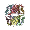 1b9lS S: Starting model for refinement |
|---|---|
| Similar structure data |

-Links
 PDBj
PDBj
- Assembly
| Deposited unit | 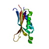
| ||||||||
|---|---|---|---|---|---|---|---|---|---|
| 1 | x 8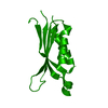
| ||||||||
| Unit cell |
|
-Components
| #1: Protein | Mass: 18011.758 Da / Num. of mol.: 1 Source method: isolated from a genetically manipulated source Source: (gene. exp.) PSEUDOMONAS AERUGINOSA (bacteria) / Production host: |
|---|---|
| #2: Water | ChemComp-HOH /  Mass: 18.015 Da / Num. of mol.: 8 / Source method: isolated from a natural source / Formula: H2O Mass: 18.015 Da / Num. of mol.: 8 / Source method: isolated from a natural source / Formula: H2O |
| Sequence details | THE PROTEIN HAS AN N-TERMINAL HIS-TAG AND TEV CLEAVAGE SITE |
-Experimental details
-Experiment
| Experiment | Method: X-RAY DIFFRACTION / Number of used crystals: 1 |
|---|
- Sample preparation
| Crystal | Density Matthews: 2.78 Å3/Da / Density % sol: 55.78 % / Description: NONE |
|---|---|
| Crystal grow | Details: 40 % (V/V) 1,2-PROPANEDIOL, 100 MM HEPES PH 7.5 |
-Data collection
| Diffraction | Mean temperature: 100 K |
|---|---|
| Diffraction source | Source: SYNCHROTRON / Site: Diamond  / Beamline: I03 / Wavelength: 0.97625 / Beamline: I03 / Wavelength: 0.97625 |
| Detector | Type: DECTRIS PILATUS 6M / Detector: PIXEL / Date: Feb 20, 2011 |
| Radiation | Protocol: SINGLE WAVELENGTH / Monochromatic (M) / Laue (L): M / Scattering type: x-ray |
| Radiation wavelength | Wavelength: 0.97625 Å / Relative weight: 1 |
| Reflection | Resolution: 3→97.74 Å / Num. obs: 4383 / % possible obs: 100 % / Observed criterion σ(I): 2.5 / Redundancy: 23.7 % / Biso Wilson estimate: 123.56 Å2 / Rmerge(I) obs: 0.05 / Net I/σ(I): 34.3 |
| Reflection shell | Resolution: 3→3.16 Å / Redundancy: 25.1 % / Rmerge(I) obs: 1.37 / Mean I/σ(I) obs: 2.9 / % possible all: 100 |
- Processing
| Software |
| ||||||||||||||||||||||||||||||||||||||||||||||||||||||||||||||||||||||||||||||||||||||||||||||||||||||||||||||||||
|---|---|---|---|---|---|---|---|---|---|---|---|---|---|---|---|---|---|---|---|---|---|---|---|---|---|---|---|---|---|---|---|---|---|---|---|---|---|---|---|---|---|---|---|---|---|---|---|---|---|---|---|---|---|---|---|---|---|---|---|---|---|---|---|---|---|---|---|---|---|---|---|---|---|---|---|---|---|---|---|---|---|---|---|---|---|---|---|---|---|---|---|---|---|---|---|---|---|---|---|---|---|---|---|---|---|---|---|---|---|---|---|---|---|---|---|
| Refinement | Method to determine structure: MOLECULAR REPLACEMENT Starting model: PDB ENTRY 1B9L Resolution: 3→66.99 Å / Cor.coef. Fo:Fc: 0.9047 / Cor.coef. Fo:Fc free: 0.878 / SU R Cruickshank DPI: 0.64 / Cross valid method: THROUGHOUT / σ(F): 0 / SU R Blow DPI: 0.703 / SU Rfree Blow DPI: 0.393 / SU Rfree Cruickshank DPI: 0.388 Details: RESIDUES 1-5 AND 47-54 ARE DISORDERED. IDEAL-DIST CONTACT TERM CONTACT SETUP. ALL ATOMS HAVE CCP4 ATOM 1B9L_MONO_CHAINSAW1.PDB
| ||||||||||||||||||||||||||||||||||||||||||||||||||||||||||||||||||||||||||||||||||||||||||||||||||||||||||||||||||
| Displacement parameters | Biso mean: 133.92 Å2
| ||||||||||||||||||||||||||||||||||||||||||||||||||||||||||||||||||||||||||||||||||||||||||||||||||||||||||||||||||
| Refinement step | Cycle: LAST / Resolution: 3→66.99 Å
| ||||||||||||||||||||||||||||||||||||||||||||||||||||||||||||||||||||||||||||||||||||||||||||||||||||||||||||||||||
| Refine LS restraints |
| ||||||||||||||||||||||||||||||||||||||||||||||||||||||||||||||||||||||||||||||||||||||||||||||||||||||||||||||||||
| LS refinement shell | Resolution: 3→3.35 Å / Total num. of bins used: 5
| ||||||||||||||||||||||||||||||||||||||||||||||||||||||||||||||||||||||||||||||||||||||||||||||||||||||||||||||||||
| Refinement TLS params. | Method: refined / Origin x: 12.9782 Å / Origin y: 13.4566 Å / Origin z: 52.8251 Å
| ||||||||||||||||||||||||||||||||||||||||||||||||||||||||||||||||||||||||||||||||||||||||||||||||||||||||||||||||||
| Refinement TLS group | Selection details: (CHAIN A) |
