 Movie
Movie Controller


+ Open data

- Basic information
| Entry | Database: PDB / ID: 4ccf | ||||||
|---|---|---|---|---|---|---|---|
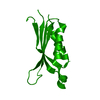
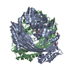
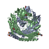
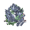
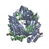
| Title | Structure of Respiratory Syncytial Virus F protein head domain | ||||||
 Components Components | FUSION GLYCOPROTEIN F0 | ||||||
 Keywords Keywords | VIRAL PROTEIN | ||||||
| Function / homology |  Function and homology information Function and homology informationsymbiont-mediated induction of syncytium formation / Translation of respiratory syncytial virus mRNAs / RSV-host interactions / Assembly and release of respiratory syncytial virus (RSV) virions / Maturation of hRSV A proteins / Respiratory syncytial virus (RSV) attachment and entry / host cell Golgi membrane / entry receptor-mediated virion attachment to host cell / fusion of virus membrane with host plasma membrane / viral envelope ...symbiont-mediated induction of syncytium formation / Translation of respiratory syncytial virus mRNAs / RSV-host interactions / Assembly and release of respiratory syncytial virus (RSV) virions / Maturation of hRSV A proteins / Respiratory syncytial virus (RSV) attachment and entry / host cell Golgi membrane / entry receptor-mediated virion attachment to host cell / fusion of virus membrane with host plasma membrane / viral envelope / symbiont entry into host cell / host cell plasma membrane / virion membrane / membrane / identical protein binding / plasma membrane Similarity search - Function | ||||||
| Biological species |  RESPIRATORY SYNCYTIAL VIRUS RESPIRATORY SYNCYTIAL VIRUS | ||||||
| Method |  X-RAY DIFFRACTION / SYNCHROTRON / MOLECULAR REPLACEMENT / Resolution: 2.65 Å X-RAY DIFFRACTION / SYNCHROTRON / MOLECULAR REPLACEMENT / Resolution: 2.65 Å | ||||||
 Authors Authors | Peat, T.S. | ||||||
 Citation Citation | Journal: To be Published Title: Structure of Respiratory Syncytial Virus F Protein Head Domain Authors: Peat, T.S. | ||||||
| History |
|
- Structure visualization
| Structure viewer | Molecule: MolmilJmol/JSmol |
|---|
- Downloads & links
-Download
| PDBx/mmCIF format | 4ccf.cif.gz | 330.6 KB | Display | PDBx/mmCIF format |
|---|---|---|---|---|
| PDB format | pdb4ccf.ent.gz | 258.1 KB | Display | PDB format |
| PDBx/mmJSON format | 4ccf.json.gz | Tree view | PDBx/mmJSON format | |
| Others |  Other downloads Other downloads |
-Validation report
| Arichive directory | https://data.pdbj.org/pub/pdb/validation_reports/cc/4ccfftp://data.pdbj.org/pub/pdb/validation_reports/cc/4ccf | HTTPS FTP |
|---|
-Related structure data
| Related structure data | 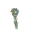 3rrrS S: Starting model for refinement |
|---|---|
| Similar structure data |

-Links
 PDBj
PDBj

- Assembly
| Deposited unit | 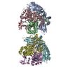
| ||||||||
|---|---|---|---|---|---|---|---|---|---|
| 1 | 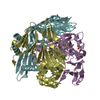
| ||||||||
| 2 | 
| ||||||||
| Unit cell |
|
-Components
| #1: Protein | Mass: 63462.969 Da / Num. of mol.: 6 / Source method: isolated from a natural source Details: THE PROTEIN WAS PROTEOLYZED BY CELLULAR PROTEASES SOMETIME DURING EXPRESSION OR PURIFICATION Source: (natural) RESPIRATORY SYNCYTIAL VIRUS / References: UniProt: G8EJ09, UniProt: P03420*PLUS#2: Chemical |   Mass: 96.063 Da / Num. of mol.: 3 / Source method: obtained synthetically / Formula: SO4 Mass: 96.063 Da / Num. of mol.: 3 / Source method: obtained synthetically / Formula: SO4#3: Water | ChemComp-HOH / |  Mass: 18.015 Da / Num. of mol.: 36 / Source method: isolated from a natural source / Formula: H2O Mass: 18.015 Da / Num. of mol.: 36 / Source method: isolated from a natural source / Formula: H2OHas protein modification | Y | Sequence details | THE PROTEIN WAS PROTEOLYZE | |
|---|
-Experimental details
-Experiment
| Experiment | Method: X-RAY DIFFRACTION / Number of used crystals: 1 |
|---|
- Sample preparation
| Crystal | Density Matthews: 3.05 Å3/Da / Density % sol: 59.6 % / Description: NONE |
|---|---|
| Crystal grow | Details: 25% PEG 8000, 200 MM AMMONIUM SULFATE, 200 MM SODIUM CACODYLATE PH 6.5 |
-Data collection
| Diffraction | Mean temperature: 100 K |
|---|---|
| Diffraction source | Source: SYNCHROTRON / Site: ESRF  / Type: ESRF / Wavelength: 1 / Type: ESRF / Wavelength: 1 |
| Detector | Type: MARRESEARCH / Detector: CCD / Date: Jun 2, 2003 |
| Radiation | Protocol: SINGLE WAVELENGTH / Monochromatic (M) / Laue (L): M / Scattering type: x-ray |
| Radiation wavelength | Wavelength: 1 Å / Relative weight: 1 |
| Reflection | Resolution: 2.65→43.1 Å / Num. obs: 53253 / % possible obs: 88 % / Observed criterion σ(I): 1 / Redundancy: 1.7 % / Rmerge(I) obs: 0.11 / Net I/σ(I): 6.2 |
| Reflection shell | Resolution: 2.65→2.79 Å / Redundancy: 1.7 % / Rmerge(I) obs: 0.67 / Mean I/σ(I) obs: 1.5 / % possible all: 85.4 |
- Processing
| Software |
| ||||||||||||||||||||||||||||||||||||||||||||||||||||||||||||||||||||||||||||||||||||||||||||||||||||||||||||||||||||||||||||||||||||||||||||||||||||||||||||||||||||||||||||||||||||||
|---|---|---|---|---|---|---|---|---|---|---|---|---|---|---|---|---|---|---|---|---|---|---|---|---|---|---|---|---|---|---|---|---|---|---|---|---|---|---|---|---|---|---|---|---|---|---|---|---|---|---|---|---|---|---|---|---|---|---|---|---|---|---|---|---|---|---|---|---|---|---|---|---|---|---|---|---|---|---|---|---|---|---|---|---|---|---|---|---|---|---|---|---|---|---|---|---|---|---|---|---|---|---|---|---|---|---|---|---|---|---|---|---|---|---|---|---|---|---|---|---|---|---|---|---|---|---|---|---|---|---|---|---|---|---|---|---|---|---|---|---|---|---|---|---|---|---|---|---|---|---|---|---|---|---|---|---|---|---|---|---|---|---|---|---|---|---|---|---|---|---|---|---|---|---|---|---|---|---|---|---|---|---|---|
| Refinement | Method to determine structure: MOLECULAR REPLACEMENT Starting model: PDB ENTRY 3RRR Resolution: 2.65→43.07 Å / Cor.coef. Fo:Fc: 0.918 / Cor.coef. Fo:Fc free: 0.888 / SU B: 12.277 / SU ML: 0.252 / Cross valid method: THROUGHOUT / ESU R: 1.746 / ESU R Free: 0.343 / Stereochemistry target values: MAXIMUM LIKELIHOOD Details: HYDROGENS HAVE BEEN ADDED IN THE RIDING POSITIONS. U VALUES REFINED INDIVIDUALLY
| ||||||||||||||||||||||||||||||||||||||||||||||||||||||||||||||||||||||||||||||||||||||||||||||||||||||||||||||||||||||||||||||||||||||||||||||||||||||||||||||||||||||||||||||||||||||
| Solvent computation | Ion probe radii: 0.8 Å / Shrinkage radii: 0.8 Å / VDW probe radii: 1.2 Å / Solvent model: MASK | ||||||||||||||||||||||||||||||||||||||||||||||||||||||||||||||||||||||||||||||||||||||||||||||||||||||||||||||||||||||||||||||||||||||||||||||||||||||||||||||||||||||||||||||||||||||
| Displacement parameters | Biso mean: 40.065 Å2
| ||||||||||||||||||||||||||||||||||||||||||||||||||||||||||||||||||||||||||||||||||||||||||||||||||||||||||||||||||||||||||||||||||||||||||||||||||||||||||||||||||||||||||||||||||||||
| Refinement step | Cycle: LAST / Resolution: 2.65→43.07 Å
| ||||||||||||||||||||||||||||||||||||||||||||||||||||||||||||||||||||||||||||||||||||||||||||||||||||||||||||||||||||||||||||||||||||||||||||||||||||||||||||||||||||||||||||||||||||||
| Refine LS restraints |
|