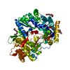
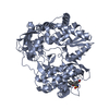
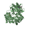
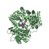
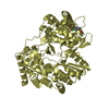
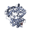
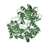
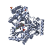
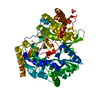
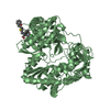
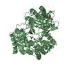
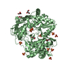
Entry Database : PDB / ID : 4adpTitle HCV-J6 NS5B POLYMERASE V405I MUTANT RNA-DIRECTED RNA POLYMERASE Keywords / / / / / / / Function / homology Function Domain/homology Component
/ / / / / / / / / / / / / / / / / / / / / / / / / / / / / / / / / / / / / / / / / / / / / / / / / / / / / / / / / / / / / / / / / / / / / / / / / / / / / / / / / / / / / / / / / / / / / / / / / / / / / / / / / / / / Biological species Method / / / Resolution : 1.902 Å Authors Scrima, N. / Bressanelli, S. Journal : J.Virol. / Year : 2012Title : Two Crucial Early Steps in RNA Synthesis by the Hepatitis C Virus Polymerase Involve a Dual Role of Residue 405.Authors : Scrima, N. / Caillet-Saguy, C. / Ventura, M. / Harrus, D. / Astier-Gin, T. / Bressanelli, S. History Deposition Jan 2, 2012 Deposition site / Processing site Revision 1.0 May 2, 2012 Provider / Type Revision 1.1 Jul 25, 2012 Group Revision 1.2 Dec 20, 2023 Group Data collection / Database references ... Data collection / Database references / Other / Refinement description Category chem_comp_atom / chem_comp_bond ... chem_comp_atom / chem_comp_bond / database_2 / pdbx_database_status / pdbx_initial_refinement_model Item / _database_2.pdbx_database_accession / _pdbx_database_status.status_code_sf
Show all Show less
 Movie
Movie Controller


 Open data
 Basic information
 Components
 Keywords
 Function and homology information
 HEPATITIS C VIRUS
 X-RAY DIFFRACTION /
  Authors
 Citation
 Structure visualization
 Downloads & links
 Other downloads


 PDBj
PDBj
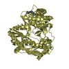
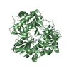
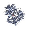
 Assembly
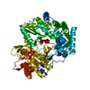

 Mass: 18.015 Da / Num. of mol.: 592 / Source method: isolated from a natural source / Formula: H2O
 Sample preparation
 / Beamline: PROXIMA 1 / Wavelength: 0.98011
  Processing