 Movie
Movie Controller

[English] 日本語
 Yorodumi
- PDB-4a65: Crystal structure of the thioredoxin reductase from Entamoeba his... -
+ Open data

- Basic information
| Entry | Database: PDB / ID: 4a65 | ||||||
|---|---|---|---|---|---|---|---|
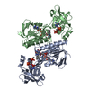
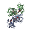
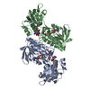
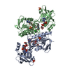
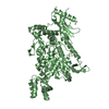
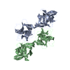
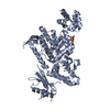
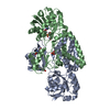
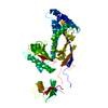
| Title | Crystal structure of the thioredoxin reductase from Entamoeba histolytica with AuCN | ||||||
 Components Components | THIOREDOXIN REDUCTASE | ||||||
 Keywords Keywords | OXIDOREDUCTASE / REDOX METABOLISM / OXIDATIVE STRESS | ||||||
| Function / homology |  Function and homology information Function and homology informationthioredoxin-disulfide reductase (NADPH) / thioredoxin-disulfide reductase (NADPH) activity / removal of superoxide radicals / cell redox homeostasis / nucleotide binding / cytosol Similarity search - Function | ||||||
| Biological species |   ENTAMOEBA HISTOLYTICA (eukaryote) ENTAMOEBA HISTOLYTICA (eukaryote) | ||||||
| Method |  X-RAY DIFFRACTION / SYNCHROTRON / MOLECULAR REPLACEMENT / Resolution: 1.7 Å X-RAY DIFFRACTION / SYNCHROTRON / MOLECULAR REPLACEMENT / Resolution: 1.7 Å | ||||||
 Authors Authors | Podust, L.M. | ||||||
 Citation Citation | Journal: J.Struct.Biol. / Year: 2016 Title: X-Ray Structures of Thioredoxin and Thioredoxin Reductase from Entamoeba Histolytica and Prevailing Hypothesis of the Mechanism of Auranofin Action. Authors: Parsonage, D. / Sheng, F. / Hirata, K. / Debnath, A. / Mckerrow, J.H. / Reed, S.L. / Abagyan, R. / Poole, L.B. / Podust, L.M. | ||||||
| History |
|
- Structure visualization
| Structure viewer | Molecule: MolmilJmol/JSmol |
|---|
- Downloads & links
-Download
| PDBx/mmCIF format | 4a65.cif.gz | 155.7 KB | Display | PDBx/mmCIF format |
|---|---|---|---|---|
| PDB format | pdb4a65.ent.gz | 121.2 KB | Display | PDB format |
| PDBx/mmJSON format | 4a65.json.gz | Tree view | PDBx/mmJSON format | |
| Others |  Other downloads Other downloads |
-Validation report
| Arichive directory | https://data.pdbj.org/pub/pdb/validation_reports/a6/4a65ftp://data.pdbj.org/pub/pdb/validation_reports/a6/4a65 | HTTPS FTP |
|---|
-Related structure data
| Related structure data |  4a5lSC  4cbqC  4ccqC  4ccrC  4cw9C  4up3C S: Starting model for refinement C: citing same article ( |
|---|---|
| Similar structure data |

-Links
 PDBj
PDBj


- Assembly
| Deposited unit | 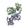
| ||||||||
|---|---|---|---|---|---|---|---|---|---|
| 1 |
| ||||||||
| Unit cell |
|
-Components
-Protein , 1 types, 2 molecules AB
| #1: Protein | Mass: 33779.598 Da / Num. of mol.: 2 Source method: isolated from a genetically manipulated source Source: (gene. exp.) ENTAMOEBA HISTOLYTICA (eukaryote) / Plasmid: PTRCHISA / Production host:  References: UniProt: C4LW95, thioredoxin-disulfide reductase (NADPH) |
|---|
-Non-polymers , 5 types, 721 molecules 


| #2: Chemical |  Mass: 745.421 Da / Num. of mol.: 2 / Source method: obtained synthetically / Formula: C21H30N7O17P3 Mass: 745.421 Da / Num. of mol.: 2 / Source method: obtained synthetically / Formula: C21H30N7O17P3#3: Chemical |  Mass: 196.967 Da / Num. of mol.: 2 / Source method: obtained synthetically / Formula: Au Mass: 196.967 Da / Num. of mol.: 2 / Source method: obtained synthetically / Formula: Au#4: Chemical |  Mass: 785.550 Da / Num. of mol.: 2 / Source method: obtained synthetically / Formula: C27H33N9O15P2 / Comment: FAD*YM Mass: 785.550 Da / Num. of mol.: 2 / Source method: obtained synthetically / Formula: C27H33N9O15P2 / Comment: FAD*YM#5: Chemical |  Mass: 96.063 Da / Num. of mol.: 2 / Source method: obtained synthetically / Formula: SO4 Mass: 96.063 Da / Num. of mol.: 2 / Source method: obtained synthetically / Formula: SO4#6: Water | ChemComp-HOH / | Mass: 18.015 Da / Num. of mol.: 713 / Source method: isolated from a natural source / Formula: H2O |
|---|
-Details
| Has protein modification | Y |
|---|---|
| Nonpolymer details | GOLD(I) (AU): BOUND TO CYS 286 |
-Experimental details
-Experiment
| Experiment | Method: X-RAY DIFFRACTION / Number of used crystals: 1 |
|---|
- Sample preparation
| Crystal | Density Matthews: 2.3 Å3/Da / Density % sol: 46.7 % / Description: NONE |
|---|---|
| Crystal grow | pH: 9 Details: 22% PEG 4000, 0.1 M BICINE PH 9.0, 0.1 M AMMONIUM SULFATE, 1 MM AUCN |
-Data collection
| Diffraction | Mean temperature: 110 K |
|---|---|
| Diffraction source | Source: SYNCHROTRON / Site: ALS  / Beamline: 8.3.1 / Wavelength: 1.11587 / Beamline: 8.3.1 / Wavelength: 1.11587 |
| Detector | Type: MARRESEARCH / Detector: CCD / Date: Oct 7, 2011 / Details: MIRRORS |
| Radiation | Monochromator: SI (111) / Protocol: SINGLE WAVELENGTH / Monochromatic (M) / Laue (L): M / Scattering type: x-ray |
| Radiation wavelength | Wavelength: 1.11587 Å / Relative weight: 1 |
| Reflection | Resolution: 1.7→101.27 Å / Num. obs: 51502 / % possible obs: 76.6 % / Observed criterion σ(I): 1.5 / Redundancy: 4.3 % / Biso Wilson estimate: 25.2 Å2 / Rmerge(I) obs: 0.05 / Net I/σ(I): 13.2 |
| Reflection shell | Resolution: 1.7→1.79 Å / Redundancy: 3.3 % / Rmerge(I) obs: 0.44 / Mean I/σ(I) obs: 2 / % possible all: 28.8 |
- Processing
| Software |
| ||||||||||||||||||||||||||||||||||||||||||||||||||||||||||||||||||||||||||||||||||||||||||||||||||||||||||||||||||||||||||||||||||||||||||||||||||||||||||||||||||||||||||||||||||||||
|---|---|---|---|---|---|---|---|---|---|---|---|---|---|---|---|---|---|---|---|---|---|---|---|---|---|---|---|---|---|---|---|---|---|---|---|---|---|---|---|---|---|---|---|---|---|---|---|---|---|---|---|---|---|---|---|---|---|---|---|---|---|---|---|---|---|---|---|---|---|---|---|---|---|---|---|---|---|---|---|---|---|---|---|---|---|---|---|---|---|---|---|---|---|---|---|---|---|---|---|---|---|---|---|---|---|---|---|---|---|---|---|---|---|---|---|---|---|---|---|---|---|---|---|---|---|---|---|---|---|---|---|---|---|---|---|---|---|---|---|---|---|---|---|---|---|---|---|---|---|---|---|---|---|---|---|---|---|---|---|---|---|---|---|---|---|---|---|---|---|---|---|---|---|---|---|---|---|---|---|---|---|---|---|
| Refinement | Method to determine structure: MOLECULAR REPLACEMENT Starting model: PDB ENTRY 4A5L Resolution: 1.7→101.27 Å / Cor.coef. Fo:Fc: 0.964 / Cor.coef. Fo:Fc free: 0.936 / SU B: 2.541 / SU ML: 0.084 / Cross valid method: THROUGHOUT / ESU R: 0.15 / ESU R Free: 0.15 / Stereochemistry target values: MAXIMUM LIKELIHOOD / Details: HYDROGENS HAVE BEEN ADDED IN THE RIDING POSITIONS.
| ||||||||||||||||||||||||||||||||||||||||||||||||||||||||||||||||||||||||||||||||||||||||||||||||||||||||||||||||||||||||||||||||||||||||||||||||||||||||||||||||||||||||||||||||||||||
| Solvent computation | Ion probe radii: 0.8 Å / Shrinkage radii: 0.8 Å / VDW probe radii: 1.4 Å / Solvent model: MASK | ||||||||||||||||||||||||||||||||||||||||||||||||||||||||||||||||||||||||||||||||||||||||||||||||||||||||||||||||||||||||||||||||||||||||||||||||||||||||||||||||||||||||||||||||||||||
| Displacement parameters | Biso mean: 25.247 Å2
| ||||||||||||||||||||||||||||||||||||||||||||||||||||||||||||||||||||||||||||||||||||||||||||||||||||||||||||||||||||||||||||||||||||||||||||||||||||||||||||||||||||||||||||||||||||||
| Refinement step | Cycle: LAST / Resolution: 1.7→101.27 Å
| ||||||||||||||||||||||||||||||||||||||||||||||||||||||||||||||||||||||||||||||||||||||||||||||||||||||||||||||||||||||||||||||||||||||||||||||||||||||||||||||||||||||||||||||||||||||
| Refine LS restraints |
|