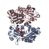
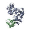
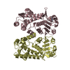
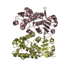
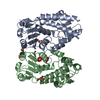
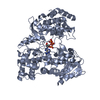
Entry Database : PDB / ID : 4a5oTitle Crystal structure of Pseudomonas aeruginosa N5, N10- methylenetetrahydrofolate dehydrogenase-cyclohydrolase (FolD) BIFUNCTIONAL PROTEIN FOLD Keywords / Function / homology Function Domain/homology Component
/ / / / / / / / / / / / / / / / / / / / / / / / / / / / / / Biological species PSEUDOMONAS AERUGINOSA PAO1 (bacteria)Method / / / Resolution : 2.2 Å Authors Eadsforth, T.C. / Gardiner, M. / Maluf, F.V. / McElroy, S. / James, D. / Frearson, J. / Gray, D. / Hunter, W.N. Journal : Plos One / Year : 2012Title : Assessment of Pseudomonas Aeruginosa N(5),N(10)-Methylenetetrahydrofolate Dehydrogenase - Cyclohydrolase as a Potential Antibacterial Drug Target.Authors : Eadsforth, T.C. / Gardiner, M. / Maluf, F.V. / Mcelroy, S. / James, D. / Frearson, J. / Gray, D. / Hunter, W.N. History Deposition Oct 26, 2011 Deposition site / Processing site Revision 1.0 Nov 16, 2011 Provider / Type Revision 1.1 May 16, 2012 Group Revision 1.2 Dec 20, 2023 Group Data collection / Database references ... Data collection / Database references / Derived calculations / Other / Refinement description Category chem_comp_atom / chem_comp_bond ... chem_comp_atom / chem_comp_bond / database_2 / pdbx_database_status / pdbx_initial_refinement_model / struct_site Item _database_2.pdbx_DOI / _database_2.pdbx_database_accession ... _database_2.pdbx_DOI / _database_2.pdbx_database_accession / _pdbx_database_status.status_code_sf / _struct_site.pdbx_auth_asym_id / _struct_site.pdbx_auth_comp_id / _struct_site.pdbx_auth_seq_id
Show all Show less
 Movie
Movie Controller

 Yorodumi
 Open data
 Basic information
 Components
 Keywords
 Function and homology information
 PSEUDOMONAS AERUGINOSA PAO1 (bacteria)
 X-RAY DIFFRACTION /
  Authors
 Citation
 Structure visualization
 Downloads & links
 Other downloads


 PDBj
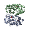
PDBj Assembly
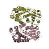
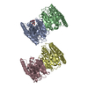

 Mass: 92.094 Da / Num. of mol.: 1 / Source method: obtained synthetically / Formula: C3H8O3
 Mass: 106.120 Da / Num. of mol.: 1 / Source method: obtained synthetically / Formula: C4H10O3
 Mass: 18.015 Da / Num. of mol.: 135 / Source method: isolated from a natural source / Formula: H2O
 Sample preparation
 / Beamline: ID29 / Wavelength: 0.9814
  Processing