Protocol: SINGLE WAVELENGTH / Monochromatic (M) / Laue (L): M / Scattering type: x-ray
Radiation wavelength
Wavelength: 0.992 Å / Relative weight: 1
Reflection
Resolution: 2.1→30.7 Å / Num. obs: 62865 / % possible obs: 99.8 % / Observed criterion σ(I): 2 / Redundancy: 6.86 % / Biso Wilson estimate: 26.95 Å2 / Rmerge(I) obs: 0.08 / Net I/σ(I): 22.99
Reflection shell
Resolution: 2.1→2.22 Å / Redundancy: 6.87 % / Rmerge(I) obs: 0.31 / Mean I/σ(I) obs: 5.18 / % possible all: 99.2
-
Processing
Software
Name
Version
Classification
BUSTER
2.8.0
refinement
SHELXCD
phasing
Refinement
Method to determine structure: SAD Starting model: NONE Resolution: 2.1→30.75 Å / Cor.coef. Fo:Fc: 0.9404 / Cor.coef. Fo:Fc free: 0.919 / Cross valid method: THROUGHOUT / σ(F): 0 Details: IDEAL-DIST CONTACT TERM CONTACT SETUP. ALL ATOMS HAVE CCP4 ATOM TYPE FROM LIBRARY. RESTRAINT LIBRARIES. NUMBER OF LIBRARIES USED: 7
Rfactor
Num. reflection
% reflection
Selection details
Rfree
0.2194
3136
5 %
RANDOM
Rwork
0.1846
-
-
-
obs
0.1863
62736
-
-
Displacement parameters
Biso mean: 32.52 Å2
Baniso -1
Baniso -2
Baniso -3
1-
-2.9536 Å2
0 Å2
0 Å2
2-
-
0.6468 Å2
0 Å2
3-
-
-
2.3067 Å2
Refine analyze
Luzzati coordinate error obs: 0.235 Å
Refinement step
Cycle: LAST / Resolution: 2.1→30.75 Å
Protein
Nucleic acid
Ligand
Solvent
Total
Num. atoms
7304
0
0
585
7889
Refine LS restraints
Refine-ID
Type
Dev ideal
Number
Restraint function
Weight
X-RAY DIFFRACTION
t_bond_d
0.01
7467
HARMONIC
2
X-RAY DIFFRACTION
t_angle_deg
1.03
10116
HARMONIC
2
X-RAY DIFFRACTION
t_dihedral_angle_d
2709
SINUSOIDAL
2
X-RAY DIFFRACTION
t_incorr_chiral_ct
X-RAY DIFFRACTION
t_pseud_angle
X-RAY DIFFRACTION
t_trig_c_planes
218
HARMONIC
2
X-RAY DIFFRACTION
t_gen_planes
1044
HARMONIC
5
X-RAY DIFFRACTION
t_it
7467
HARMONIC
20
X-RAY DIFFRACTION
t_nbd
X-RAY DIFFRACTION
t_omega_torsion
2.76
X-RAY DIFFRACTION
t_other_torsion
17.31
X-RAY DIFFRACTION
t_improper_torsion
X-RAY DIFFRACTION
t_chiral_improper_torsion
979
SEMIHARMONIC
5
X-RAY DIFFRACTION
t_sum_occupancies
X-RAY DIFFRACTION
t_utility_distance
X-RAY DIFFRACTION
t_utility_angle
X-RAY DIFFRACTION
t_utility_torsion
X-RAY DIFFRACTION
t_ideal_dist_contact
9450
SEMIHARMONIC
4
LS refinement shell
Resolution: 2.1→2.15 Å / Total num. of bins used: 20
Rfactor
Num. reflection
% reflection
Rfree
0.1992
227
4.99 %
Rwork
0.1864
4321
-
all
0.187
4548
-
Refinement TLS params.
Method: refined / Refine-ID: X-RAY DIFFRACTION
ID
L11 (°2)
L12 (°2)
L13 (°2)
L22 (°2)
L23 (°2)
L33 (°2)
S11 (Å °)
S12 (Å °)
S13 (Å °)
S21 (Å °)
S22 (Å °)
S23 (Å °)
S31 (Å °)
S32 (Å °)
S33 (Å °)
T11 (Å2)
T12 (Å2)
T13 (Å2)
T22 (Å2)
T23 (Å2)
T33 (Å2)
Origin x (Å)
Origin y (Å)
Origin z (Å)
1
0.2263
0.0244
-0.1781
0.6219
-0.4616
0.5041
0.0277
-0.0271
-0.0106
0.0179
-0.0621
-0.0492
-0.0202
0.0786
0.0343
-0.0357
-0.0043
-0.0221
-0.0393
-0.0232
-0.0582
32.5155
2.5991
0.3563
2
0.1966
0.1136
0.2018
0.3862
0.3302
0.4805
0.0222
-0.0411
0.0106
0.0558
-0.0287
0.0196
0.0294
-0.0919
0.0065
-0.0456
0.0023
0.0283
-0.0223
0.0113
-0.0419
49.2979
-2.976
-1.6073
Refinement TLS group
ID
Refine-ID
Refine TLS-ID
Selection details
1
X-RAY DIFFRACTION
1
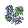
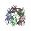
CHAIN A AND RESID 8-21 OR RESID 27-42 OR RESID 51-64 OR RESID 72-418 OR RESID 424-465
2
X-RAY DIFFRACTION
2
CHAIN B AND RESID 7-465
+
About Yorodumi
-
News
-
Feb 9, 2022. New format data for meta-information of EMDB entries
New format data for meta-information of EMDB entries
Version 3 of the EMDB header file is now the official format.
The previous official version 1.9 will be removed from the archive.
In the structure databanks used in Yorodumi, some data are registered as the other names, "COVID-19 virus" and "2019-nCoV". Here are the details of the virus and the list of structure data.
Jan 31, 2019. EMDB accession codes are about to change! (news from PDBe EMDB page)
EMDB accession codes are about to change! (news from PDBe EMDB page)
The allocation of 4 digits for EMDB accession codes will soon come to an end. Whilst these codes will remain in use, new EMDB accession codes will include an additional digit and will expand incrementally as the available range of codes is exhausted. The current 4-digit format prefixed with “EMD-” (i.e. EMD-XXXX) will advance to a 5-digit format (i.e. EMD-XXXXX), and so on. It is currently estimated that the 4-digit codes will be depleted around Spring 2019, at which point the 5-digit format will come into force.
The EM Navigator/Yorodumi systems omit the EMD- prefix.
Related info.:Q: What is EMD? / ID/Accession-code notation in Yorodumi/EM Navigator
Yorodumi is a browser for structure data from EMDB, PDB, SASBDB, etc.
This page is also the successor to EM Navigator detail page, and also detail information page/front-end page for Omokage search.
The word "yorodu" (or yorozu) is an old Japanese word meaning "ten thousand". "mi" (miru) is to see.
Related info.:EMDB / PDB / SASBDB / Comparison of 3 databanks / Yorodumi Search / Aug 31, 2016. New EM Navigator & Yorodumi / Yorodumi Papers / Jmol/JSmol / Function and homology information / Changes in new EM Navigator and Yorodumi
 Movie
Movie Controller


 Open data
 Basic information
 Components
 Keywords
 Function and homology information
 X-RAY DIFFRACTION /
  Authors
 Citation
 Structure visualization
 Downloads & links
 Other downloads
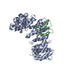


 PDBj
PDBj
 Assembly
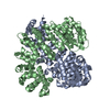

 Mass: 18.015 Da / Num. of mol.: 585 / Source method: isolated from a natural source / Formula: H2O
 Sample preparation
 / Beamline: PROXIMA 1 / Wavelength: 0.992
  Processing