 Movie
Movie Controller

[English] 日本語
 Yorodumi
- PDB-4a3h: 2',4' DINITROPHENYL-2-DEOXY-2-FLURO-B-D-CELLOBIOSIDE COMPLEX OF T... -
+ Open data

- Basic information
| Entry | Database: PDB / ID: 4a3h | ||||||
|---|---|---|---|---|---|---|---|
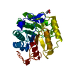
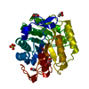
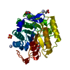
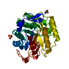
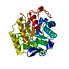
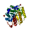
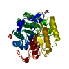
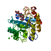
| Title | 2',4' DINITROPHENYL-2-DEOXY-2-FLURO-B-D-CELLOBIOSIDE COMPLEX OF THE ENDOGLUCANASE CEL5A FROM BACILLUS AGARADHAERENS AT 1.6 A RESOLUTION | ||||||
 Components Components | PROTEIN (ENDOGLUCANASE) | ||||||
 Keywords Keywords | HYDROLASE / CELLULOSE DEGRADATION / ENDOGLUCANASE / GLYCOSIDE HYDROLASE FAMILY 5 / MICHAELIS COMPLEX. SKEW-BOAT / DISTORTION | ||||||
| Function / homology |  Function and homology information Function and homology informationcellulase / cellulase activity / cellulose catabolic process / carbohydrate binding / extracellular region Similarity search - Function | ||||||
| Biological species |  Bacillus agaradhaerens (bacteria) Bacillus agaradhaerens (bacteria) | ||||||
| Method |  X-RAY DIFFRACTION / ISOMORPHOUS WITH NATIVE STRUCTURE / Resolution: 1.65 Å X-RAY DIFFRACTION / ISOMORPHOUS WITH NATIVE STRUCTURE / Resolution: 1.65 Å | ||||||
 Authors Authors | Davies, G.J. / Brzozowski, A.M. / Andersen, K. / Schulein, M. / Mackenzie, L. / Withers, S.G. | ||||||
 Citation Citation | Journal: Biochemistry / Year: 1998 Title: Snapshots along an enzymatic reaction coordinate: analysis of a retaining beta-glycoside hydrolase. Authors: Davies, G.J. / Mackenzie, L. / Varrot, A. / Dauter, M. / Brzozowski, A.M. / Schulein, M. / Withers, S.G. #1: Journal: Biochemistry / Year: 1998Title: Structure of the Bacillus Agaradherans Family 5 Endoglucanase at 1.6 A and its Cellobiose Complex at 2.0 A Resolution Authors: Davies, G.J. / Dauter, M. / Brzozowski, A.M. / Bjornvad, M.E. / Andersen, K.V. / Schulein, M. | ||||||
| History |
|
- Structure visualization
| Structure viewer | Molecule: MolmilJmol/JSmol |
|---|
- Downloads & links
-Download
| PDBx/mmCIF format | 4a3h.cif.gz | 82.1 KB | Display | PDBx/mmCIF format |
|---|---|---|---|---|
| PDB format | pdb4a3h.ent.gz | 61.4 KB | Display | PDB format |
| PDBx/mmJSON format | 4a3h.json.gz | Tree view | PDBx/mmJSON format | |
| Others |  Other downloads Other downloads |
-Validation report
| Arichive directory | https://data.pdbj.org/pub/pdb/validation_reports/a3/4a3hftp://data.pdbj.org/pub/pdb/validation_reports/a3/4a3h | HTTPS FTP |
|---|
-Related structure data


-Links
 PDBj
PDBj- Assembly
| Deposited unit | 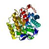
| ||||||||
|---|---|---|---|---|---|---|---|---|---|
| 1 |
| ||||||||
| Unit cell |
|
-Components
| #1: Protein | Mass: 33998.023 Da / Num. of mol.: 1 / Fragment: CATALYTIC CORE DOMAIN ONLY Source method: isolated from a genetically manipulated source Details: THIS IS A COMPLEX WITH 2,4-DINITROPHENYL, 2-FLUOROCELLOBIOSEOUND IN THE -1, -2 AND +1 SITES OF THE ENZYME Source: (gene. exp.) Bacillus agaradhaerens (bacteria) / Strain: AC13 (NCIMB 40482) / Plasmid: THERMAMYL-AMYLASE PROMOTER SYSTEM / Production host: References: UniProt: P06565, UniProt: O85465*PLUS, cellulase | ||
|---|---|---|---|
| #2: Chemical | ChemComp-DCB /   Mass: 510.379 Da / Num. of mol.: 1 / Source method: obtained synthetically / Formula: C18H23FN2O14 Mass: 510.379 Da / Num. of mol.: 1 / Source method: obtained synthetically / Formula: C18H23FN2O14 | ||
| #3: Water | ChemComp-HOH /  Mass: 18.015 Da / Num. of mol.: 417 / Source method: isolated from a natural source / Formula: H2O Mass: 18.015 Da / Num. of mol.: 417 / Source method: isolated from a natural source / Formula: H2O | ||
| Compound details | CEL5A IS A MEMBER OF GLYCOSIDE HYDROLASE FAMILY 5 THIS IS ONE OF THE GH-A CLAN MEMBERS | ||
| Nonpolymer details | HET ID DCB THE CELLOBIOSE| Sequence details | REFERENCE: SEQUENCE NOT DEPOSITED. | |
-Experimental details
-Experiment
| Experiment | Method: X-RAY DIFFRACTION / Number of used crystals: 1 |
|---|
- Sample preparation
| Crystal | Density Matthews: 2.16 Å3/Da / Density % sol: 42.94 % | |||||||||||||||
|---|---|---|---|---|---|---|---|---|---|---|---|---|---|---|---|---|
| Crystal grow | pH: 5.5 Details: PROTEIN (20MGML-1) WAS CRYSTALLISED FROM 1.0M AMMONIUM SULPHATE AS BOTH BUFFER AND PRECIPITANT AT PH 4.5 IN THE PRESENCE OF 15% (W/V) GLYCEROL, pH 5.5 THIS STRUCTURE WAS OBTAINED BY SOAKING ...Details: PROTEIN (20MGML-1) WAS CRYSTALLISED FROM 1.0M AMMONIUM SULPHATE AS BOTH BUFFER AND PRECIPITANT AT PH 4.5 IN THE PRESENCE OF 15% (W/V) GLYCEROL, pH 5.5 THIS STRUCTURE WAS OBTAINED BY SOAKING THE CRYSTALS IN 10MM 2",4" DINITROPHENYL-2-DEOXY-2-FLUORO-B-D-CELLOBIOSIDE FOR 12 H PRIOR TO DATA COLLECTION. | |||||||||||||||
| Crystal grow | *PLUS pH: 4.5 / Method: vapor diffusion, hanging drop / Details: Davies, G.J., (1998) Biochemistry, 37, 1926. | |||||||||||||||
| Components of the solutions | *PLUS
|
-Data collection
| Diffraction | Mean temperature: 100 K |
|---|---|
| Diffraction source | Source: ROTATING ANODE / Type: RIGAKU RU200 / Wavelength: 1.5418 |
| Detector | Type: MARRESEARCH / Detector: IMAGE PLATE / Date: May 15, 1997 / Details: LONG FOCUSSING MIRRORS (MSC) |
| Radiation | Protocol: SINGLE WAVELENGTH / Monochromatic (M) / Laue (L): M / Scattering type: x-ray |
| Radiation wavelength | Wavelength: 1.5418 Å / Relative weight: 1 |
| Reflection | Resolution: 1.65→20 Å / Num. obs: 35964 / % possible obs: 99.9 % / Redundancy: 4.2 % / Biso Wilson estimate: 14.6 Å2 / Rmerge(I) obs: 0.046 / Rsym value: 0.046 / Net I/σ(I): 22.6 |
| Reflection shell | Resolution: 1.65→1.71 Å / Redundancy: 4.2 % / Rmerge(I) obs: 0.176 / Mean I/σ(I) obs: 13.4 / Rsym value: 0.176 / % possible all: 99.9 |
| Reflection shell | *PLUS % possible obs: 99.8 % |
- Processing
| Software |
| ||||||||||||||||||||||||||||||||||||||||||||||||||||||||||||||||||||||||||||||||||||
|---|---|---|---|---|---|---|---|---|---|---|---|---|---|---|---|---|---|---|---|---|---|---|---|---|---|---|---|---|---|---|---|---|---|---|---|---|---|---|---|---|---|---|---|---|---|---|---|---|---|---|---|---|---|---|---|---|---|---|---|---|---|---|---|---|---|---|---|---|---|---|---|---|---|---|---|---|---|---|---|---|---|---|---|---|---|
| Refinement | Method to determine structure: ISOMORPHOUS WITH NATIVE STRUCTURE Resolution: 1.65→15 Å / Cross valid method: THROUGHOUT / σ(F): 0
| ||||||||||||||||||||||||||||||||||||||||||||||||||||||||||||||||||||||||||||||||||||
| Displacement parameters | Biso mean: 15.8 Å2 | ||||||||||||||||||||||||||||||||||||||||||||||||||||||||||||||||||||||||||||||||||||
| Refinement step | Cycle: LAST / Resolution: 1.65→15 Å
| ||||||||||||||||||||||||||||||||||||||||||||||||||||||||||||||||||||||||||||||||||||
| Refine LS restraints |
| ||||||||||||||||||||||||||||||||||||||||||||||||||||||||||||||||||||||||||||||||||||
| Software | *PLUS Name: REFMAC / Classification: refinement | ||||||||||||||||||||||||||||||||||||||||||||||||||||||||||||||||||||||||||||||||||||
| Refinement | *PLUS Lowest resolution: 15 Å / σ(F): 0 / % reflection Rfree: 5 % / Rfactor obs: 0.14 | ||||||||||||||||||||||||||||||||||||||||||||||||||||||||||||||||||||||||||||||||||||
| Solvent computation | *PLUS | ||||||||||||||||||||||||||||||||||||||||||||||||||||||||||||||||||||||||||||||||||||
| Displacement parameters | *PLUS Biso mean: 15.8 Å2 | ||||||||||||||||||||||||||||||||||||||||||||||||||||||||||||||||||||||||||||||||||||
| Refine LS restraints | *PLUS
|
