 Movie
Movie Controller

[English] 日本語
 Yorodumi
- PDB-1hf6: ENDOGLUCANASE CEL5A FROM BACILLUS AGARADHAERENS IN THE ORTHORHOMB... -
+ Open data

- Basic information
| Entry | Database: PDB / ID: 1hf6 | |||||||||
|---|---|---|---|---|---|---|---|---|---|---|
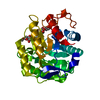
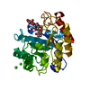
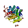
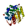
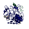
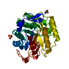
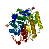
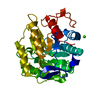
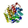
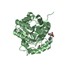
| Title | ENDOGLUCANASE CEL5A FROM BACILLUS AGARADHAERENS IN THE ORTHORHOMBIC CRYSTAL FORM IN COMPLEX WITH CELLOTRIOSE | |||||||||
 Components Components | ENDOGLUCANASE B | |||||||||
 Keywords Keywords | HYDROLASE / CELLULOSE DEGRADATION / ENDOGLUCANASE / GLYCOSHYDROLASE FAMILY 5 | |||||||||
| Function / homology |  Function and homology information Function and homology informationcellulase / cellulase activity / cellulose catabolic process / carbohydrate binding / extracellular region Similarity search - Function | |||||||||
| Biological species |  BACILLUS AGARADHAERENS (bacteria) BACILLUS AGARADHAERENS (bacteria) | |||||||||
| Method |  X-RAY DIFFRACTION / SYNCHROTRON / MOLECULAR REPLACEMENT / Resolution: 1.15 Å X-RAY DIFFRACTION / SYNCHROTRON / MOLECULAR REPLACEMENT / Resolution: 1.15 Å | |||||||||
 Authors Authors | Varrot, A. / Withers, S. / Vasella, A. / Schulein, M. / Davies, G.J. | |||||||||
 Citation Citation | Journal: Acta Crystallogr.,Sect.D / Year: 2003 Title: Direct Experimental Observation of the Hydrogen-Bonding Network of a Glycosidase Along its Reaction Coordinate Revealed by Atomic Resolution Analyses of Endoglucanase Cel5A Authors: Varrot, A. / Davies, G.J. | |||||||||
| History |
| |||||||||
| Remark 700 | SHEET DETERMINATION METHOD: DSSP THE SHEETS PRESENTED AS "AB" IN EACH CHAIN ON SHEET RECORDS BELOW ... SHEET DETERMINATION METHOD: DSSP THE SHEETS PRESENTED AS "AB" IN EACH CHAIN ON SHEET RECORDS BELOW IS ACTUALLY AN 8-STRANDED BARREL THIS IS REPRESENTED BY A 9-STRANDED SHEET IN WHICH THE FIRST AND LAST STRANDS ARE IDENTICAL. |
- Structure visualization
| Structure viewer | Molecule: MolmilJmol/JSmol |
|---|
- Downloads & links
-Download
| PDBx/mmCIF format | 1hf6.cif.gz | 156.5 KB | Display | PDBx/mmCIF format |
|---|---|---|---|---|
| PDB format | pdb1hf6.ent.gz | 122.7 KB | Display | PDB format |
| PDBx/mmJSON format | 1hf6.json.gz | Tree view | PDBx/mmJSON format | |
| Others |  Other downloads Other downloads |
-Validation report
| Arichive directory | https://data.pdbj.org/pub/pdb/validation_reports/hf/1hf6ftp://data.pdbj.org/pub/pdb/validation_reports/hf/1hf6 | HTTPS FTP |
|---|
-Related structure data
| Related structure data |  1h11C 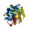 1h2jC  3a3hS C: citing same article ( S: Starting model for refinement |
|---|---|
| Similar structure data |


-Links
 PDBj
PDBj

- Assembly
| Deposited unit | 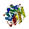
| ||||||||
|---|---|---|---|---|---|---|---|---|---|
| 1 |
| ||||||||
| Unit cell |
| ||||||||
| Details | BIOLOGICAL_UNIT: ACTIVE AS A MONOMER |
-Components
-Protein / Sugars , 2 types, 2 molecules A
| #1: Protein | Mass: 33998.023 Da / Num. of mol.: 1 / Fragment: CATALYTIC CORE DOMAIN ONLY Source method: isolated from a genetically manipulated source Source: (gene. exp.) BACILLUS AGARADHAERENS (bacteria) / Plasmid: THERMAMYL-AMYLASE PROMOT / Production host: |
|---|---|
| #2: Polysaccharide | beta-D-glucopyranose-(1-4)-beta-D-glucopyranose-(1-4)-alpha-D-glucopyranose / alpha-cellotriose  Source method: isolated from a genetically manipulated source Details: oligosaccharide / References: alpha-cellotriose |
-Non-polymers , 4 types, 475 molecules 


| #3: Chemical | ChemComp-SO4 /  Mass: 96.063 Da / Num. of mol.: 1 / Source method: obtained synthetically / Formula: SO4 Mass: 96.063 Da / Num. of mol.: 1 / Source method: obtained synthetically / Formula: SO4 | ||
|---|---|---|---|
| #4: Chemical | ChemComp-ACY /  Mass: 60.052 Da / Num. of mol.: 1 / Source method: obtained synthetically / Formula: C2H4O2 Mass: 60.052 Da / Num. of mol.: 1 / Source method: obtained synthetically / Formula: C2H4O2 | ||
| #5: Chemical |  Mass: 92.094 Da / Num. of mol.: 2 / Source method: obtained synthetically / Formula: C3H8O3 Mass: 92.094 Da / Num. of mol.: 2 / Source method: obtained synthetically / Formula: C3H8O3#6: Water | ChemComp-HOH / | Mass: 18.015 Da / Num. of mol.: 471 / Source method: isolated from a natural source / Formula: H2O |
-Details
| Sequence details | THE FIRST 26 RESIDUES IN THE DATABASE CORRESPOND TO THE PROSEQUENCE. OUR NUMBERING BEGIN AT THE ...THE FIRST 26 RESIDUES IN THE DATABASE CORRESPOND |
|---|
-Experimental details
-Experiment
| Experiment | Method: X-RAY DIFFRACTION / Number of used crystals: 1 |
|---|
- Sample preparation
| Crystal | Density Matthews: 1.9 Å3/Da / Density % sol: 35.58 % | |||||||||||||||
|---|---|---|---|---|---|---|---|---|---|---|---|---|---|---|---|---|
| Crystal grow | pH: 4.6 Details: PROTEIN CONCENTRATION 20MG/ML, 2M AMMONIUM SULPHATE, 100MM SODIUM CITRATE PH 5.5, 25% GLYCEROL AS CRYOPROTECTANT | |||||||||||||||
| Crystal grow | *PLUS pH: 4.5 / Method: vapor diffusion, hanging drop / Details: Davies, G.J., (1998) Biochemistry, 37, 1926. | |||||||||||||||
| Components of the solutions | *PLUS
|
-Data collection
| Diffraction | Mean temperature: 100 K |
|---|---|
| Diffraction source | Source: SYNCHROTRON / Site: EMBL/DESY, HAMBURG  / Beamline: X31 / Wavelength: 1.069 / Beamline: X31 / Wavelength: 1.069 |
| Detector | Type: MARRESEARCH / Detector: IMAGE PLATE / Date: Oct 15, 1998 |
| Radiation | Protocol: SINGLE WAVELENGTH / Monochromatic (M) / Laue (L): M / Scattering type: x-ray |
| Radiation wavelength | Wavelength: 1.069 Å / Relative weight: 1 |
| Reflection | Resolution: 1.15→20 Å / Num. obs: 102353 / % possible obs: 98.9 % / Redundancy: 4.3 % / Biso Wilson estimate: 7.5 Å2 / Rmerge(I) obs: 0.053 / Rsym value: 0.053 / Net I/σ(I): 24.8 |
| Reflection shell | Resolution: 1.15→1.19 Å / Redundancy: 3.4 % / Rmerge(I) obs: 0.307 / Mean I/σ(I) obs: 3.9 / Rsym value: 0.307 / % possible all: 93 |
| Reflection | *PLUS Highest resolution: 1.15 Å / Lowest resolution: 20 Å / Redundancy: 4.3 % |
| Reflection shell | *PLUS % possible obs: 93 % / Redundancy: 3.4 % / Mean I/σ(I) obs: 3.9 |
- Processing
| Software |
| ||||||||||||||||||||
|---|---|---|---|---|---|---|---|---|---|---|---|---|---|---|---|---|---|---|---|---|---|
| Refinement | Method to determine structure: MOLECULAR REPLACEMENT Starting model: PDB ENTRY 3A3H Resolution: 1.15→20 Å / SU B: 0.955 / SU ML: 0.023 / Cross valid method: THROUGHOUT / ESU R: 0.043 / ESU R Free: 0.041 / Stereochemistry target values: MAXIMUM LIKELIHOOD Details: HYDROGENS HAVE BEEN ADDED IN THE RIDING POSITIONS. THE FIRST THREE RESIDUES WERE NOT VISIBLE IN DENSITY
| ||||||||||||||||||||
| Displacement parameters | Biso mean: 9.17 Å2
| ||||||||||||||||||||
| Refinement step | Cycle: LAST / Resolution: 1.15→20 Å
| ||||||||||||||||||||
| Refine LS restraints | *PLUS
|
