Mass: 1464.043 Da / Num. of mol.: 1 / Source method: obtained synthetically / Formula: C81H156O17P2 / Comment: phospholipid*YM
Has protein modification
N
Nonpolymer details
C9 ALKYL CHAIN (PLM): THIS IS A PUTATIVE LIGAND CONSISTING OF 8 CARBON ATOMS ARRANGED AS IN (3R, ...C9 ALKYL CHAIN (PLM): THIS IS A PUTATIVE LIGAND CONSISTING OF 8 CARBON ATOMS ARRANGED AS IN (3R, 12R)-3-AMINO-12- METHYLTETRADECANAL. COMPLETE CHEMICAL INFORMATION IS NOT KNOWN
-
Experimental details
-
Experiment
Experiment
Method: X-RAY DIFFRACTION / Number of used crystals: 1
-
Sample preparation
Crystal
Density Matthews: 7.15 Å3/Da / Density % sol: 80.2 % / Description: NONE
Crystal grow
pH: 6.5 Details: 0.1 M AMMONIUM SULPHATE, 0.3 M SODIUM FORMATE, 0.1 M SODIUM CACODYLATE PH 6.5, 3% (V/V) POLYGLUTAMIC ACID AND 20%-26% (W/V) PEG 550 MME
Protocol: SINGLE WAVELENGTH / Monochromatic (M) / Laue (L): M / Scattering type: x-ray
Radiation wavelength
Wavelength: 0.939 Å / Relative weight: 1
Reflection
Resolution: 3.4→47.54 Å / Num. obs: 9625 / % possible obs: 98.3 % / Observed criterion σ(I): 1 / Redundancy: 6.7 % / Biso Wilson estimate: 97.06 Å2 / Rmerge(I) obs: 0.13 / Net I/σ(I): 9.3
Reflection shell
Resolution: 3.4→3.58 Å / Redundancy: 7 % / Rmerge(I) obs: 0.86 / Mean I/σ(I) obs: 2.1 / % possible all: 99
-
Processing
Software
Name
Classification
MOSFLM
datareduction
SCALA
datascaling
PHENIX
phasing
PHASER
phasing
PHENIX
refinement
Refinement
Method to determine structure: SAD Starting model: NONE Resolution: 3.4→38.947 Å / SU ML: 0.5 / σ(F): 1.02 / Phase error: 25.86 / Stereochemistry target values: MLHL Details: SIDE CHAIN ATOMS FOR WHICH ELECTRON DENSITY IS NOT OBSERVED ARE NOT INCLUDED IN THE REFINEMENT.
Rfactor
Num. reflection
% reflection
Rfree
0.2749
462
4.9 %
Rwork
0.2384
-
-
obs
0.24
9625
98.2 %
Solvent computation
Shrinkage radii: 0.49 Å / VDW probe radii: 0.8 Å / Solvent model: FLAT BULK SOLVENT MODEL / Bsol: 54.518 Å2 / ksol: 0.278 e/Å3
Displacement parameters
Biso mean: 108.81 Å2
Baniso -1
Baniso -2
Baniso -3
1-
-13.2349 Å2
0 Å2
0 Å2
2-
-
-13.2349 Å2
0 Å2
3-
-
-
26.4697 Å2
Refinement step
Cycle: LAST / Resolution: 3.4→38.947 Å
Protein
Nucleic acid
Ligand
Solvent
Total
Num. atoms
1554
0
58
0
1612
Refine LS restraints
Refine-ID
Type
Dev ideal
Number
X-RAY DIFFRACTION
f_bond_d
0.01
1658
X-RAY DIFFRACTION
f_angle_d
1.341
2255
X-RAY DIFFRACTION
f_dihedral_angle_d
19.687
583
X-RAY DIFFRACTION
f_chiral_restr
0.087
254
X-RAY DIFFRACTION
f_plane_restr
0.005
267
LS refinement shell
Resolution (Å)
Rfactor Rfree
Num. reflection Rfree
Rfactor Rwork
Num. reflection Rwork
Refine-ID
% reflection obs (%)
3.4-3.6129
0.3503
151
0.3517
2680
X-RAY DIFFRACTION
99
3.6129-3.8916
0.3343
147
0.2847
2683
X-RAY DIFFRACTION
99
3.8916-4.2828
0.2496
121
0.2394
2712
X-RAY DIFFRACTION
99
4.2828-4.9015
0.2652
143
0.1952
2675
X-RAY DIFFRACTION
99
4.9015-6.1714
0.2665
125
0.2292
2692
X-RAY DIFFRACTION
98
6.1714-38.9499
0.2477
136
0.2245
2636
X-RAY DIFFRACTION
96
Refinement TLS params.
Method: refined / Refine-ID: X-RAY DIFFRACTION
ID
L11 (°2)
L12 (°2)
L13 (°2)
L22 (°2)
L23 (°2)
L33 (°2)
S11 (Å °)
S12 (Å °)
S13 (Å °)
S21 (Å °)
S22 (Å °)
S23 (Å °)
S31 (Å °)
S32 (Å °)
S33 (Å °)
T11 (Å2)
T12 (Å2)
T13 (Å2)
T22 (Å2)
T23 (Å2)
T33 (Å2)
Origin x (Å)
Origin y (Å)
Origin z (Å)
1
0.4268
0.408
-0.1113
0.4029
-0.0403
0.2649
-0.1487
1.2458
-1.0515
-0.1288
-0.1411
-0.0993
0.1774
-0.7084
0.0694
0.5827
0.0674
0.0151
1.2472
0.0728
0.8519
21.4014
44.4165
84.2142
2
0.3138
-0.2699
0.5077
0.214
-0.4212
0.7436
-0.0845
0.4476
-0.4133
0.1404
-0.085
-0.5519
-0.0532
-0.2328
0.1477
0.3178
0.1148
0.0093
0.9447
-0.0257
0.481
12.9466
48.672
95.0824
3
0.0582
0.0467
-0.1613
0.1953
-0.3747
0.8259
0.0707
0.2199
-0.3562
-0.1834
0.4313
0.7337
0.4025
-0.7846
-0.0916
0.843
0.151
-0.0922
1.4567
0.1963
0.736
8.1103
49.499
115.8241
4
0.6097
-0.4047
-0.0625
0.2591
0.0095
0.1566
-0.3042
0.1243
-0.007
0.1286
-0.2281
-0.0687
-0.0575
0.1776
0.075
0.1908
-0.1058
0.0013
0.9475
0.0793
0.3618
11.478
56.9625
95.3641
5
0.2007
0.1286
-0.4699
0.7035
0.2983
1.6486
-0.7291
-0.9226
0.5451
0.7688
-0.188
-0.2337
0.2972
-1.4023
-0.0406
0.7798
0.2384
-0.1206
0.8183
-0.2589
0.5946
5.9067
61.8217
114.462
6
0.5278
0.1191
0.2478
0.2671
-0.0754
0.2425
-0.3874
-0.8836
0.6659
-0.16
-0.2633
-0.7323
0.3625
-0.3104
0.0682
0.8042
0.0422
0.1165
0.8348
-0.1585
1.204
13.8855
67.6874
106.3112
Refinement TLS group
ID
Refine-ID
Refine TLS-ID
Selection details
1
X-RAY DIFFRACTION
1
(CHAINBANDRESID1:18)
2
X-RAY DIFFRACTION
2
(CHAINBANDRESID19:107)
3
X-RAY DIFFRACTION
3
(CHAINBANDRESID108:113)
4
X-RAY DIFFRACTION
4
(CHAINBANDRESID114:166)
5
X-RAY DIFFRACTION
5
(CHAINBANDRESID167:180)
6
X-RAY DIFFRACTION
6
(CHAINBANDRESID181:192)
+
About Yorodumi
-
News
-
Feb 9, 2022. New format data for meta-information of EMDB entries
New format data for meta-information of EMDB entries
Version 3 of the EMDB header file is now the official format.
The previous official version 1.9 will be removed from the archive.
In the structure databanks used in Yorodumi, some data are registered as the other names, "COVID-19 virus" and "2019-nCoV". Here are the details of the virus and the list of structure data.
Jan 31, 2019. EMDB accession codes are about to change! (news from PDBe EMDB page)
EMDB accession codes are about to change! (news from PDBe EMDB page)
The allocation of 4 digits for EMDB accession codes will soon come to an end. Whilst these codes will remain in use, new EMDB accession codes will include an additional digit and will expand incrementally as the available range of codes is exhausted. The current 4-digit format prefixed with “EMD-” (i.e. EMD-XXXX) will advance to a 5-digit format (i.e. EMD-XXXXX), and so on. It is currently estimated that the 4-digit codes will be depleted around Spring 2019, at which point the 5-digit format will come into force.
The EM Navigator/Yorodumi systems omit the EMD- prefix.
Related info.:Q: What is EMD? / ID/Accession-code notation in Yorodumi/EM Navigator
Yorodumi is a browser for structure data from EMDB, PDB, SASBDB, etc.
This page is also the successor to EM Navigator detail page, and also detail information page/front-end page for Omokage search.
The word "yorodu" (or yorozu) is an old Japanese word meaning "ten thousand". "mi" (miru) is to see.
Related info.:EMDB / PDB / SASBDB / Comparison of 3 databanks / Yorodumi Search / Aug 31, 2016. New EM Navigator & Yorodumi / Yorodumi Papers / Jmol/JSmol / Function and homology information / Changes in new EM Navigator and Yorodumi
 Movie
Movie Controller


 Open data
 Basic information
 Components
 Keywords
 Function and homology information
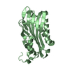
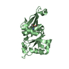
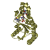
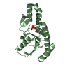
 METHANOSARCINA ACETIVORANS (archaea)
 X-RAY DIFFRACTION /
  Authors
 Citation
 Structure visualization
 Downloads & links
 Other downloads


 PDBj
PDBj


 Assembly


 Mass: 384.411 Da / Num. of mol.: 1 / Source method: obtained synthetically / Formula: C14H20N6O5S
 Mass: 256.424 Da / Num. of mol.: 1 / Source method: obtained synthetically / Formula: C16H32O2
 Mass: 1464.043 Da / Num. of mol.: 1 / Source method: obtained synthetically / Formula: C81H156O17P2 / Comment: phospholipid*YM
 Sample preparation
 / Beamline: I03 / Wavelength: 0.939
  Processing