 Movie
Movie Controller


+ Open data

- Basic information
| Entry | Database: PDB / ID: 452d | ||||||||||||||||||
|---|---|---|---|---|---|---|---|---|---|---|---|---|---|---|---|---|---|---|---|
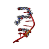
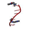
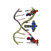
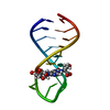
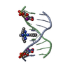
| Title | ACRIDINE BINDING TO DNA | ||||||||||||||||||
 Components Components | DNA (5'-D(* Keywords KeywordsDNA / INTERCALATION / GROOVE BINDING | Function / homology | Chem-9AD / DNA |  Function and homology information Function and homology informationMethod |  X-RAY DIFFRACTION / Resolution: 1.6 Å X-RAY DIFFRACTION / Resolution: 1.6 Å  Authors AuthorsThorpe, J.H. / Todd, A.K. / Cardin, C.J. |  CitationJournal: J.Med.Chem. / Year: 1999 CitationJournal: J.Med.Chem. / Year: 1999Title: Major groove binding and 'DNA-induced' fit in the intercalation of a derivative of the mixed topoisomerase I/II poison N-(2-(dimethlyamino)ethyl)acridine-4-carboxamide (DACA) into DNA: X-ray ...Title: Major groove binding and 'DNA-induced' fit in the intercalation of a derivative of the mixed topoisomerase I/II poison N-(2-(dimethlyamino)ethyl)acridine-4-carboxamide (DACA) into DNA: X-ray structure complexed to d(CG(5Br-U)ACG)2 at 1.3-angstrom resolution Authors: Todd, A.K. / Adams, A. / Thorpe, J.H. / Denny, W.A. / Wakelin, L.P.G. / Cardin, C.J. History |
|
- Structure visualization
| Structure viewer | Molecule: MolmilJmol/JSmol |
|---|
- Downloads & links
-Download
| PDBx/mmCIF format | 452d.cif.gz | 17.6 KB | Display | PDBx/mmCIF format |
|---|---|---|---|---|
| PDB format | pdb452d.ent.gz | 11 KB | Display | PDB format |
| PDBx/mmJSON format | 452d.json.gz | Tree view | PDBx/mmJSON format | |
| Others |  Other downloads Other downloads |
-Validation report
| Arichive directory | https://data.pdbj.org/pub/pdb/validation_reports/52/452dftp://data.pdbj.org/pub/pdb/validation_reports/52/452d | HTTPS FTP |
|---|
-Related structure data


-Links
 PDBj
PDBj

- Assembly
| Deposited unit | 
| ||||||||||
|---|---|---|---|---|---|---|---|---|---|---|---|
| 1 | 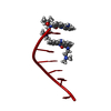
| ||||||||||
| Unit cell |
| ||||||||||
| Components on special symmetry positions |
|
-Components
| #1: DNA chain | Mass: 1809.217 Da / Num. of mol.: 1 / Source method: obtained synthetically | ||||
|---|---|---|---|---|---|
| #2: Chemical |   Mass: 308.378 Da / Num. of mol.: 2 / Source method: obtained synthetically / Formula: C18H20N4O Mass: 308.378 Da / Num. of mol.: 2 / Source method: obtained synthetically / Formula: C18H20N4O#3: Chemical | ChemComp-MPD / ( |   Mass: 118.174 Da / Num. of mol.: 1 / Source method: obtained synthetically / Formula: C6H14O2 / Comment: precipitant*YM Mass: 118.174 Da / Num. of mol.: 1 / Source method: obtained synthetically / Formula: C6H14O2 / Comment: precipitant*YM#4: Water | ChemComp-HOH / |  Mass: 18.015 Da / Num. of mol.: 29 / Source method: isolated from a natural source / Formula: H2O Mass: 18.015 Da / Num. of mol.: 29 / Source method: isolated from a natural source / Formula: H2O |
-Experimental details
-Experiment
| Experiment | Method: X-RAY DIFFRACTION / Number of used crystals: 1 |
|---|
- Sample preparation
| Crystal | Density Matthews: 2.76 Å3/Da / Density % sol: 55.41 % |
|---|---|
| Crystal grow | Method: vapor diffusion, sitting drop / Details: VAPOR DIFFUSION, SITTING DROP |
-Data collection
| Diffraction source | Source: ROTATING ANODE / Wavelength: 1.5418 |
|---|---|
| Detector | Detector: AREA DETECTOR |
| Radiation | Protocol: SINGLE WAVELENGTH / Monochromatic (M) / Laue (L): M / Scattering type: x-ray |
| Radiation wavelength | Wavelength: 1.5418 Å / Relative weight: 1 |
| Reflection | Resolution: 1.6→16 Å |
- Processing
| Software | Name: SHELXL-97 / Classification: refinement | ||||||||||||
|---|---|---|---|---|---|---|---|---|---|---|---|---|---|
| Refinement | Resolution: 1.6→16 Å
| ||||||||||||
| Refinement step | Cycle: LAST / Resolution: 1.6→16 Å
|
