 Movie
Movie Controller


+ Open data

- Basic information
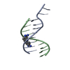
| Entry | Database: PDB / ID: 431d | ||||||||||||||||||
|---|---|---|---|---|---|---|---|---|---|---|---|---|---|---|---|---|---|---|---|
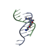
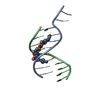
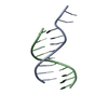
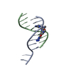
| Title | 5'-D(*GP*GP*CP*CP*AP*AP*TP*TP*GP*G)-3' | ||||||||||||||||||
 Components Components | DNA (5'-D(* Keywords KeywordsDNA / DEOXYRIBONUCLEIC ACID / EXTENDED HYDRATION SPINE / TRIPLET FORMATION / ATOMIC RESOLUTION / DOUBLE BACKBONE CONFORMATION | Function / homology | DNA |  Function and homology information Function and homology informationMethod |  X-RAY DIFFRACTION / SYNCHROTRON / Resolution: 1.15 Å X-RAY DIFFRACTION / SYNCHROTRON / Resolution: 1.15 Å  Authors AuthorsVlieghe, D. / Turkenburg, J.P. / Van Meervelt, L. |  CitationJournal: Acta Crystallogr.,Sect.D / Year: 1999 CitationJournal: Acta Crystallogr.,Sect.D / Year: 1999Title: B-DNA at atomic resolution reveals extended hydration patterns. Authors: Vlieghe, D. / Turkenburg, J.P. / Van Meervelt, L. #1: Journal: Science / Year: 1996Title: Parallel and Antiparallel (G-Gc)2 Triple Helix Fragments in a Crystal Structure Authors: Vlieghe, D. / Van Meervelt, L. / Dautant, A. / Gallois, B. / Precigoux, G. / Kennard, O. History |
|
- Structure visualization
| Structure viewer | Molecule: MolmilJmol/JSmol |
|---|
- Downloads & links
-Download
| PDBx/mmCIF format | 431d.cif.gz | 37.1 KB | Display | PDBx/mmCIF format |
|---|---|---|---|---|
| PDB format | pdb431d.ent.gz | 26.2 KB | Display | PDB format |
| PDBx/mmJSON format | 431d.json.gz | Tree view | PDBx/mmJSON format | |
| Others |  Other downloads Other downloads |
-Validation report
| Arichive directory | https://data.pdbj.org/pub/pdb/validation_reports/31/431dftp://data.pdbj.org/pub/pdb/validation_reports/31/431d | HTTPS FTP |
|---|
-Related structure data
| Similar structure data |
|---|

-Links
 PDBj
PDBj

- Assembly
| Deposited unit | 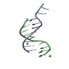
| ||||||||||
|---|---|---|---|---|---|---|---|---|---|---|---|
| 1 |
| ||||||||||
| Unit cell |
|
-Components
| #1: DNA chain | Mass: 3085.029 Da / Num. of mol.: 2 / Source method: obtained synthetically #2: Chemical |   Mass: 24.305 Da / Num. of mol.: 2 / Source method: obtained synthetically / Formula: Mg Mass: 24.305 Da / Num. of mol.: 2 / Source method: obtained synthetically / Formula: Mg#3: Water | ChemComp-HOH / |  Mass: 18.015 Da / Num. of mol.: 116 / Source method: isolated from a natural source / Formula: H2O Mass: 18.015 Da / Num. of mol.: 116 / Source method: isolated from a natural source / Formula: H2O |
|---|
-Experimental details
-Experiment
| Experiment | Method: X-RAY DIFFRACTION / Number of used crystals: 1 |
|---|
- Sample preparation
| Crystal | Density Matthews: 2.03 Å3/Da / Density % sol: 39.31 % | ||||||||||||||||||||||||||||||||||||||||||
|---|---|---|---|---|---|---|---|---|---|---|---|---|---|---|---|---|---|---|---|---|---|---|---|---|---|---|---|---|---|---|---|---|---|---|---|---|---|---|---|---|---|---|---|
| Crystal grow | Temperature: 289 K / Method: vapor diffusion, sitting drop / pH: 6 Details: pH 6.0, VAPOR DIFFUSION, SITTING DROP, temperature 289.0K | ||||||||||||||||||||||||||||||||||||||||||
| Components of the solutions |
| ||||||||||||||||||||||||||||||||||||||||||
| Crystal grow | *PLUS Temperature: 289. K / pH: 6 / Method: unknown | ||||||||||||||||||||||||||||||||||||||||||
| Components of the solutions | *PLUS
|
-Data collection
| Diffraction | Mean temperature: 120 K |
|---|---|
| Diffraction source | Source: SYNCHROTRON / Site: ELETTRA  / Beamline: 5.2R / Beamline: 5.2R |
| Detector | Type: MARRESEARCH / Detector: IMAGE PLATE / Date: May 23, 1996 |
| Radiation | Protocol: SINGLE WAVELENGTH / Monochromatic (M) / Laue (L): M / Scattering type: x-ray |
| Radiation wavelength | Relative weight: 1 |
| Reflection | Resolution: 1.15→15 Å / Num. all: 17760 / Num. obs: 17760 / % possible obs: 96.7 % / Observed criterion σ(F): 0 / Observed criterion σ(I): 0 / Redundancy: 3.6 % / Rmerge(I) obs: 0.054 / Net I/σ(I): 25 |
| Reflection shell | Resolution: 1.15→1.18 Å / Redundancy: 3.2 % / Rmerge(I) obs: 0.158 / Mean I/σ(I) obs: 11.71 / % possible all: 94.7 |
| Reflection | *PLUS Num. measured all: 63114 |
| Reflection shell | *PLUS % possible obs: 95 % |
- Processing
| Software |
| |||||||||||||||||||||||||||||||||
|---|---|---|---|---|---|---|---|---|---|---|---|---|---|---|---|---|---|---|---|---|---|---|---|---|---|---|---|---|---|---|---|---|---|---|
| Refinement | Starting model: UDJ049 USED AS STARTING MODEL FOR REFINEMENT Resolution: 1.15→10 Å / Num. parameters: 4811 / Num. restraintsaints: 12226 / Cross valid method: NONE StereochEM target val spec case: ALL OTHER CHEMICALLY EQUIVALENT BOND DISTANCES AND ANGLES WERE RESTRAINED TO BE SIMILAR BY SAME DISTANCE RESTRAINTS. Stereochemistry target values: DISTANCES AND ANGLES WITHIN THE BASES WERE RESTRAINED TO TARGET VALUES (TAYLOR AND KENNARD). Details: SIGMAS USED BOND LENGTHS (TARGET + SIMILAR) 0.03; ANGLE DISTANCES (TARGET + SIMILAR) 0.05; PLANE RESTRAINT FOR BASES 0.02; CHIRAL VOLUME RESTRAINT (A**3) 0.2; ANTI-BUMPING RESTRAINT FOR ...Details: SIGMAS USED BOND LENGTHS (TARGET + SIMILAR) 0.03; ANGLE DISTANCES (TARGET + SIMILAR) 0.05; PLANE RESTRAINT FOR BASES 0.02; CHIRAL VOLUME RESTRAINT (A**3) 0.2; ANTI-BUMPING RESTRAINT FOR SOLVENT MOLECULES 0.03; ISOR RESTRAINT FOR 28 SOLVENTS 0.1, FOR 15 SOLVENTS AND 21 DNA ATOMS 0.05, FOR 3 SOLVENTS 0.025; RIGID- BOND ADP COMPONENTS 0.010; SIMILAR ADP COMPONENTS 0.062
| |||||||||||||||||||||||||||||||||
| Solvent computation | Solvent model: MOEWS & KRETSINGER,J.MOL.BIOL.91(1973)201-222. | |||||||||||||||||||||||||||||||||
| Refine analyze | Num. disordered residues: 2 / Occupancy sum hydrogen: 527.61 / Occupancy sum non hydrogen: 208 | |||||||||||||||||||||||||||||||||
| Refinement step | Cycle: LAST / Resolution: 1.15→10 Å
| |||||||||||||||||||||||||||||||||
| Refine LS restraints |
| |||||||||||||||||||||||||||||||||
| Software | *PLUS Name: SHELXL-93 / Classification: refinement | |||||||||||||||||||||||||||||||||
| Refinement | *PLUS Lowest resolution: 10 Å / σ(F): 4 / Rfactor obs: 0.167 | |||||||||||||||||||||||||||||||||
| Solvent computation | *PLUS | |||||||||||||||||||||||||||||||||
| Displacement parameters | *PLUS | |||||||||||||||||||||||||||||||||
| Refine LS restraints | *PLUS Type: s_plane_restr / Dev ideal: 0.015 |
