 Movie
Movie Controller

[English] 日本語
 Yorodumi
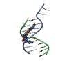
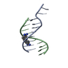
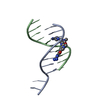
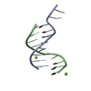
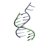
- PDB-1k2z: The Crystal Structure of d(GGCCAATTGG) Complexed with Distamycin. -
+ Open data

- Basic information
| Entry | Database: PDB / ID: 1k2z | ||||||||||||||||||
|---|---|---|---|---|---|---|---|---|---|---|---|---|---|---|---|---|---|---|---|
| Title | The Crystal Structure of d(GGCCAATTGG) Complexed with Distamycin. | ||||||||||||||||||
 Components Components | 5'-D(* Keywords KeywordsDNA / B-DNA double helix / base triplets / minor groove binding / drug / distamycin | Function / homology | DISTAMYCIN A / DNA |  Function and homology information Function and homology informationMethod |  X-RAY DIFFRACTION / SYNCHROTRON / MOLECULAR REPLACEMENT / Resolution: 2.38 Å X-RAY DIFFRACTION / SYNCHROTRON / MOLECULAR REPLACEMENT / Resolution: 2.38 Å  Authors AuthorsUytterhoeven, K. / Van Meervelt, L. |  CitationJournal: Eur.J.Biochem. / Year: 2002 CitationJournal: Eur.J.Biochem. / Year: 2002Title: Two 1 : 1 binding modes for distamycin in the minor groove of d(GGCCAATTGG). Authors: Uytterhoeven, K. / Sponer, J. / Van Meervelt, L. History |
|
- Structure visualization
| Structure viewer | Molecule: MolmilJmol/JSmol |
|---|
- Downloads & links
-Download
| PDBx/mmCIF format | 1k2z.cif.gz | 23.1 KB | Display | PDBx/mmCIF format |
|---|---|---|---|---|
| PDB format | pdb1k2z.ent.gz | 14.5 KB | Display | PDB format |
| PDBx/mmJSON format | 1k2z.json.gz | Tree view | PDBx/mmJSON format | |
| Others |  Other downloads Other downloads |
-Validation report
| Arichive directory | https://data.pdbj.org/pub/pdb/validation_reports/k2/1k2zftp://data.pdbj.org/pub/pdb/validation_reports/k2/1k2z | HTTPS FTP |
|---|
-Related structure data
| Related structure data |  1jtlC C: citing same article ( |
|---|---|
| Similar structure data | |
| Other databases |
|
-Links
 PDBj
PDBj

- Assembly
| Deposited unit | 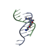
| ||||||||
|---|---|---|---|---|---|---|---|---|---|
| 1 |
| ||||||||
| Unit cell |
|
-Components
| #1: DNA chain | Mass: 3085.029 Da / Num. of mol.: 2 / Source method: obtained synthetically Details: This sequence is synthesized by Oswell DNA Service (Southampton, UK). #2: Chemical | ChemComp-DMY / | 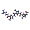  Mass: 481.508 Da / Num. of mol.: 1 / Source method: obtained synthetically / Formula: C22H27N9O4 / Comment: antibiotic*YM Mass: 481.508 Da / Num. of mol.: 1 / Source method: obtained synthetically / Formula: C22H27N9O4 / Comment: antibiotic*YM#3: Water | ChemComp-HOH / |  Mass: 18.015 Da / Num. of mol.: 46 / Source method: isolated from a natural source / Formula: H2O Mass: 18.015 Da / Num. of mol.: 46 / Source method: isolated from a natural source / Formula: H2O |
|---|
-Experimental details
-Experiment
| Experiment | Method: X-RAY DIFFRACTION / Number of used crystals: 1 |
|---|
- Sample preparation
| Crystal | Density Matthews: 2.21 Å3/Da / Density % sol: 44.31 % | ||||||||||||||||||||||||||||
|---|---|---|---|---|---|---|---|---|---|---|---|---|---|---|---|---|---|---|---|---|---|---|---|---|---|---|---|---|---|
| Crystal grow | Temperature: 293 K / Method: vapor diffusion, sitting drop / pH: 6 Details: MPD, magnesium chloride, cacodylate, spermine, pH 6.0, VAPOR DIFFUSION, SITTING DROP, temperature 293K | ||||||||||||||||||||||||||||
| Components of the solutions |
|
-Data collection
| Diffraction | Mean temperature: 100 K |
|---|---|
| Diffraction source | Source: SYNCHROTRON / Site: EMBL/DESY, HAMBURG  / Beamline: X11 / Wavelength: 0.9116 Å / Beamline: X11 / Wavelength: 0.9116 Å |
| Detector | Type: MARRESEARCH / Detector: IMAGE PLATE / Date: Apr 9, 1999 |
| Radiation | Monochromator: Si 111 / Protocol: SINGLE WAVELENGTH / Monochromatic (M) / Laue (L): M / Scattering type: x-ray |
| Radiation wavelength | Wavelength: 0.9116 Å / Relative weight: 1 |
| Reflection | Resolution: 2.38→50 Å / Num. all: 2538 / Num. obs: 2351 / % possible obs: 92.7 % / Observed criterion σ(F): 2.5 / Observed criterion σ(I): 5 / Redundancy: 5.5 % / Biso Wilson estimate: 80.64 Å2 / Rmerge(I) obs: 0.041 / Net I/σ(I): 22.6 |
| Reflection shell | Resolution: 2.38→2.42 Å / Redundancy: 3.8 % / Mean I/σ(I) obs: 5.8 / Num. unique all: 113 / Rsym value: 0.251 / % possible all: 93.4 |
- Processing
| Software |
| ||||||||||||
|---|---|---|---|---|---|---|---|---|---|---|---|---|---|
| Refinement | Method to determine structure: MOLECULAR REPLACEMENT Starting model: NDB entry DD0002 Resolution: 2.38→100 Å / Isotropic thermal model: Isotropic / σ(F): 0 / Stereochemistry target values: user defined
| ||||||||||||
| Displacement parameters | Biso mean: 72.9 Å2 | ||||||||||||
| Refinement step | Cycle: LAST / Resolution: 2.38→100 Å
| ||||||||||||
| Refine LS restraints |
|
