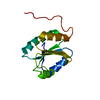
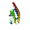
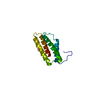
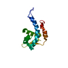
- PDB-3zyw: Crystal structure of the first glutaredoxin domain of human gluta... -
+
Open data
ID or keywords:
Loading...
-
Basic information
Entry
Database: PDB / ID: 3zyw
Title
Crystal structure of the first glutaredoxin domain of human glutaredoxin 3 (GLRX3)
Components
GLUTAREDOXIN-3
Keywords
METAL BINDING PROTEIN
Function / homology
Function and homology information
negative regulation of cardiac muscle hypertrophy / regulation of the force of heart contraction / iron-sulfur cluster assembly complex / [2Fe-2S] cluster assembly / iron-sulfur cluster assembly / iron-sulfur cluster binding / cell redox homeostasis / protein kinase C binding / Iron uptake and transport / Z disc ...negative regulation of cardiac muscle hypertrophy / regulation of the force of heart contraction / iron-sulfur cluster assembly complex / [2Fe-2S] cluster assembly / iron-sulfur cluster assembly / iron-sulfur cluster binding / cell redox homeostasis / protein kinase C binding / Iron uptake and transport / Z disc / cell cortex / intracellular iron ion homeostasis / dendrite / RNA binding / metal ion binding / identical protein binding / nucleus / cytoplasm / cytosol Similarity search - Function
GLUTAREDOXIN-3 / PKC-INTERACTING COUSIN OF THIOREDOXIN / PICOT / PKC-THETA-INTERACTING PROTEIN / PKCQ-INTERACTING ...PKC-INTERACTING COUSIN OF THIOREDOXIN / PICOT / PKC-THETA-INTERACTING PROTEIN / PKCQ-INTERACTING PROTEIN / THIOREDOXIN-LIKE PROTEIN 2
Mass: 12742.636 Da / Num. of mol.: 2 / Fragment: GLUTAREDOXIN DOMAIN, RESIDUES 130-232 Source method: isolated from a genetically manipulated source Source: (gene. exp.) HOMO SAPIENS (human) / Plasmid: PNIC-CTHF / Production host: ESCHERICHIA COLI (E. coli) / Strain (production host): BL21(DE3) / Variant (production host): R3-PRARE2 / References: UniProt: O76003
Mass: 18.015 Da / Num. of mol.: 410 / Source method: isolated from a natural source / Formula: H2O
Sequence details
STARTING MET0 IN CHAIN B AS WELL AS TERMINAL RESIDUES ALA233 GLU234 ASN235 LEU236 TYR237 PHE238 ...STARTING MET0 IN CHAIN B AS WELL AS TERMINAL RESIDUES ALA233 GLU234 ASN235 LEU236 TYR237 PHE238 GLN239 ARE DUE TO CONSTRUCT DESIGN
-
Experimental details
-
Experiment
Experiment
Method: X-RAY DIFFRACTION / Number of used crystals: 1
-
Sample preparation
Crystal
Density Matthews: 2.69 Å3/Da / Density % sol: 54.22 % / Description: NONE
Resolution: 1.84→18.95 Å / Cor.coef. Fo:Fc: 0.939 / Cor.coef. Fo:Fc free: 0.91 / SU B: 3.773 / SU ML: 0.112 / Cross valid method: THROUGHOUT / ESU R: 0.153 / ESU R Free: 0.149 / Stereochemistry target values: MAXIMUM LIKELIHOOD Details: HYDROGENS HAVE BEEN ADDED IN THE RIDING POSITIONS. U VALUES REFINED INDIVIDUALLY.
Rfactor
Num. reflection
% reflection
Selection details
Rfree
0.24974
1215
5.1 %
RANDOM
Rwork
0.19975
-
-
-
obs
0.20241
22530
97.22 %
-
Solvent computation
Ion probe radii: 0.8 Å / Shrinkage radii: 0.8 Å / VDW probe radii: 1.2 Å / Solvent model: MASK
 Movie
Movie Controller

 Yorodumi
 Open data
 Basic information
 Components
 Keywords
 Function and homology information
 HOMO SAPIENS (human)
 X-RAY DIFFRACTION /
  Authors
 Citation
 Structure visualization
 Downloads & links
 Other downloads
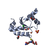


 PDBj
PDBj

 Assembly


 Mass: 62.068 Da / Num. of mol.: 5 / Source method: obtained synthetically / Formula: C2H6O2
 Mass: 18.015 Da / Num. of mol.: 410 / Source method: isolated from a natural source / Formula: H2O
 Sample preparation
 / Beamline: I03 / Wavelength: 0.9763
  Processing