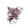
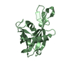
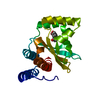
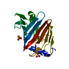
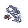
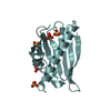
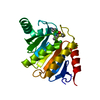
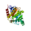
E69 deletion mutant single insulin-like growth factor binding domain protein (SIBD-1) from Cupiennius salei
Components
SINGLE INSULIN-LIKE GROWTH FACTOR-BINDING DOMAIN PROTEIN-1
Keywords
SIGNALING / IGFBP / SINGLE INSULIN-BINDING DOMAIN
Function / homology
Function and homology information
insulin-like growth factor binding / regulation of signal transduction / regulation of cell growth / innate immune response / extracellular region Similarity search - Function
Type: MARRESEARCH / Detector: CCD / Date: Jun 8, 2010
Radiation
Protocol: SINGLE WAVELENGTH / Monochromatic (M) / Laue (L): M / Scattering type: x-ray
Radiation wavelength
Wavelength: 1 Å / Relative weight: 1
Reflection
Resolution: 1.4→100 Å / Num. obs: 34438 / % possible obs: 98.6 % / Observed criterion σ(I): 2.16 / Redundancy: 5.7 % / Biso Wilson estimate: 17.76 Å2 / Rmerge(I) obs: 0.04 / Net I/σ(I): 19
Reflection shell
Resolution: 1.4→1.48 Å / Redundancy: 5.4 % / Rmerge(I) obs: 0.07 / Mean I/σ(I) obs: 2.16 / % possible all: 95.9
-
Processing
Software
Name
Version
Classification
PHENIX
(PHENIX.REFINE)
refinement
XDS
datareduction
XSCALE
datascaling
SHELXCD
phasing
SHELXE
phasing
Refinement
Method to determine structure: SAD Starting model: NONE Resolution: 1.4→35.007 Å / SU ML: 0.15 / σ(F): 1.99 / Phase error: 19.31 / Stereochemistry target values: ML Details: RESIDUES 61-66 IN CHAIN A, 64-67 IN CHAIN B ARE DISORDERED
Rfactor
Num. reflection
% reflection
Rfree
0.191
1722
5 %
Rwork
0.1594
-
-
obs
0.161
34397
98.83 %
Solvent computation
Shrinkage radii: 0.65 Å / VDW probe radii: 0.8 Å / Solvent model: FLAT BULK SOLVENT MODEL / Bsol: 52.16 Å2 / ksol: 0.457 e/Å3
Displacement parameters
Baniso -1
Baniso -2
Baniso -3
1-
1.5121 Å2
0 Å2
0 Å2
2-
-
1.5121 Å2
0 Å2
3-
-
-
-3.0242 Å2
Refinement step
Cycle: LAST / Resolution: 1.4→35.007 Å
Protein
Nucleic acid
Ligand
Solvent
Total
Num. atoms
1036
0
8
242
1286
Refine LS restraints
Refine-ID
Type
Dev ideal
Number
X-RAY DIFFRACTION
f_bond_d
0.008
1082
X-RAY DIFFRACTION
f_angle_d
1.118
1454
X-RAY DIFFRACTION
f_dihedral_angle_d
21.059
663
X-RAY DIFFRACTION
f_chiral_restr
0.061
152
X-RAY DIFFRACTION
f_plane_restr
0.006
200
LS refinement shell
Resolution (Å)
Rfactor Rfree
Num. reflection Rfree
Rfactor Rwork
Num. reflection Rwork
Refine-ID
% reflection obs (%)
1.4-1.4412
0.308
136
0.2527
2585
X-RAY DIFFRACTION
96
1.4412-1.4877
0.2548
143
0.21
2716
X-RAY DIFFRACTION
100
1.4877-1.5409
0.2025
143
0.1744
2707
X-RAY DIFFRACTION
100
1.5409-1.6026
0.2104
143
0.156
2707
X-RAY DIFFRACTION
100
1.6026-1.6755
0.2013
142
0.1444
2689
X-RAY DIFFRACTION
100
1.6755-1.7639
0.1983
144
0.14
2744
X-RAY DIFFRACTION
100
1.7639-1.8744
0.2031
144
0.146
2733
X-RAY DIFFRACTION
100
1.8744-2.0191
0.1805
144
0.1449
2726
X-RAY DIFFRACTION
100
2.0191-2.2222
0.1885
145
0.1442
2766
X-RAY DIFFRACTION
100
2.2222-2.5437
0.2132
146
0.1574
2756
X-RAY DIFFRACTION
100
2.5437-3.2045
0.1881
148
0.1603
2826
X-RAY DIFFRACTION
100
3.2045-35.0177
0.1681
144
0.1642
2720
X-RAY DIFFRACTION
92
+
About Yorodumi
-
News
-
Feb 9, 2022. New format data for meta-information of EMDB entries
New format data for meta-information of EMDB entries
Version 3 of the EMDB header file is now the official format.
The previous official version 1.9 will be removed from the archive.
In the structure databanks used in Yorodumi, some data are registered as the other names, "COVID-19 virus" and "2019-nCoV". Here are the details of the virus and the list of structure data.
Jan 31, 2019. EMDB accession codes are about to change! (news from PDBe EMDB page)
EMDB accession codes are about to change! (news from PDBe EMDB page)
The allocation of 4 digits for EMDB accession codes will soon come to an end. Whilst these codes will remain in use, new EMDB accession codes will include an additional digit and will expand incrementally as the available range of codes is exhausted. The current 4-digit format prefixed with “EMD-” (i.e. EMD-XXXX) will advance to a 5-digit format (i.e. EMD-XXXXX), and so on. It is currently estimated that the 4-digit codes will be depleted around Spring 2019, at which point the 5-digit format will come into force.
The EM Navigator/Yorodumi systems omit the EMD- prefix.
Related info.:Q: What is EMD? / ID/Accession-code notation in Yorodumi/EM Navigator
Yorodumi is a browser for structure data from EMDB, PDB, SASBDB, etc.
This page is also the successor to EM Navigator detail page, and also detail information page/front-end page for Omokage search.
The word "yorodu" (or yorozu) is an old Japanese word meaning "ten thousand". "mi" (miru) is to see.
Related info.:EMDB / PDB / SASBDB / Comparison of 3 databanks / Yorodumi Search / Aug 31, 2016. New EM Navigator & Yorodumi / Yorodumi Papers / Jmol/JSmol / Function and homology information / Changes in new EM Navigator and Yorodumi
 Movie
Movie Controller

 Yorodumi
 Open data
 Basic information
 Components
 Keywords
 Function and homology information
 CUPIENNIUS SALEI (spider)
 X-RAY DIFFRACTION /
  Authors
 Citation
 Structure visualization
 Downloads & links
 Other downloads
 PDBj
PDBj


 Assembly


 Mass: 59.044 Da / Num. of mol.: 2 / Source method: obtained synthetically / Formula: C2H3O2
 Mass: 18.015 Da / Num. of mol.: 242 / Source method: isolated from a natural source / Formula: H2O
 Sample preparation
 / Beamline: X06DA / Wavelength: 1
  Processing