- PDB-3zxb: Wild type single insulin-like growth factor binding domain protei... -
+
Open data
ID or keywords:
Loading...
-
Basic information
Entry
Database: PDB / ID: 3zxb
Title
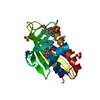
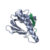
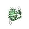
Wild type single insulin-like growth factor binding domain protein (SIBD-1) from Cupiennius salei
Components
SINGLE INSULIN-LIKE GROWTH FACTOR-BINDING DOMAIN PROTEIN-1
Keywords
SIGNALING / IGFBP / INSULIN BINDING DOMAIN
Function / homology
Function and homology information
insulin-like growth factor binding / regulation of signal transduction / regulation of cell growth / innate immune response / extracellular region Similarity search - Function
Resolution: 2.55→51.366 Å / σ(F): 1.99 / Phase error: 29.14 / Stereochemistry target values: TWIN_LSQ_F Details: RESIDUES 59-69 IN CHAIN A, 60-69 IN CHAIN B, 61-69 IN CHAIN C, 59-69 IN CHAIN D ARE DISORDERED.
Rfactor
Num. reflection
% reflection
Rfree
0.2504
653
5 %
Rwork
0.2241
-
-
obs
0.2257
13035
99.78 %
Solvent computation
Shrinkage radii: 0.95 Å / VDW probe radii: 1.2 Å / Solvent model: FLAT BULK SOLVENT MODEL / Bsol: 85.545 Å2 / ksol: 0.354 e/Å3
Displacement parameters
Baniso -1
Baniso -2
Baniso -3
1-
15.3196 Å2
0 Å2
0 Å2
2-
-
15.3196 Å2
0 Å2
3-
-
-
-30.6393 Å2
Refinement step
Cycle: LAST / Resolution: 2.55→51.366 Å
Protein
Nucleic acid
Ligand
Solvent
Total
Num. atoms
1895
0
0
41
1936
Refine LS restraints
Refine-ID
Type
Dev ideal
Number
X-RAY DIFFRACTION
f_bond_d
0.009
1971
X-RAY DIFFRACTION
f_angle_d
1.425
2648
X-RAY DIFFRACTION
f_dihedral_angle_d
20.986
1206
X-RAY DIFFRACTION
f_chiral_restr
0.079
274
X-RAY DIFFRACTION
f_plane_restr
0.011
363
LS refinement shell
Resolution (Å)
Rfactor Rfree
Num. reflection Rfree
Rfactor Rwork
Num. reflection Rwork
Refine-ID
% reflection obs (%)
2.5505-2.8072
0.3063
160
0.3053
3033
X-RAY DIFFRACTION
95
2.8072-3.2133
0.3119
159
0.2774
3027
X-RAY DIFFRACTION
95
3.2133-4.0482
0.2469
162
0.2337
3079
X-RAY DIFFRACTION
95
4.0482-51.3767
0.2252
171
0.193
3232
X-RAY DIFFRACTION
94
Refinement TLS params.
Method: refined / Refine-ID: X-RAY DIFFRACTION
ID
L11 (°2)
L12 (°2)
L13 (°2)
L22 (°2)
L23 (°2)
L33 (°2)
S11 (Å °)
S12 (Å °)
S13 (Å °)
S21 (Å °)
S22 (Å °)
S23 (Å °)
S31 (Å °)
S32 (Å °)
S33 (Å °)
T11 (Å2)
T12 (Å2)
T13 (Å2)
T22 (Å2)
T23 (Å2)
T33 (Å2)
Origin x (Å)
Origin y (Å)
Origin z (Å)
1
0.5251
0.319
1.2116
-0.0475
0.3517
3.0698
-0.6763
-0.733
0.0301
0.5087
0.2572
-0.2491
-1.8177
-0.5467
0.3031
0.9301
0.2783
0.0739
0.6441
0.1333
0.5526
-23.5237
31.6321
27.786
2
5.5082
0.4543
-3.2587
2.3885
0.1497
2.018
0.666
0.8814
-0.3003
0.0037
-0.4093
-0.1011
-0.5137
-0.7384
-0.0526
0.529
-0.0264
-0.0111
0.6741
0.0216
0.4215
-24.2382
31.5899
37.1754
3
3.0258
2.2462
0.1684
6.8453
-1.6524
1.0066
0.5275
-1.3914
0.5104
-1.1281
-0.184
0.6966
0.5243
1.0739
-0.1782
0.3963
-0.073
-0.0573
0.5026
-0.1839
0.3726
-14.4867
28.1702
40.9723
4
7.0303
-0.7687
-0.4718
3.3951
-1.1044
2.5137
-0.5101
-0.5604
0.5498
-0.3526
0.9624
-0.144
0.4452
0.0084
-0.1858
0.4574
0.0126
-0.0813
0.4689
-0.0796
0.4337
-20.9943
31.8455
49.9004
5
2.2638
1.677
-0.3832
7.5967
3.5757
3.6555
0.2222
0.4418
1.2856
0.8584
-0.0379
0.7175
1.9537
1.3515
-0.0278
0.6685
0.2424
-0.0372
0.642
0.0311
0.4975
-13.3756
19.4939
45.3468
6
3.2512
0.6737
2.5692
6.277
-1.0655
6.5541
-1.2221
0.3184
-0.6735
-0.0417
0.5192
-1.1707
-0.7253
-0.6027
0.8106
1.1982
-0.0021
0.2227
0.2847
-0.1015
0.4017
-21.2965
16.4864
32.4742
7
4.4843
0.2728
-2.0771
1.2783
0.7136
5.9238
0.8656
1.2098
0.1207
-0.2693
-0.4774
0.0173
-0.836
0.4686
-0.693
0.7458
-0.1372
0.1194
0.6083
-0.0832
0.3512
-18.7178
20.0113
29.8487
8
0.8456
-0.631
0.3778
0.5047
0.168
1.0009
-0.0837
0.0432
0.3616
-0.1504
-0.2069
-0.4126
-0.5124
-0.3061
0.1349
0.2303
0.1033
0.1401
0.6482
0.0154
0.4175
-14.0795
22.8687
22.7632
9
5.4728
2.1956
1.415
3.0944
0.3678
9.7303
-1.0444
-0.4913
-0.1715
0.0507
0.2686
1.1189
-2.8035
-2.6921
0.4592
0.9342
0.3589
0.1084
1.144
0.1415
0.5019
-9.2058
53.0647
38.5494
10
0.9185
0.0694
-0.5121
2.5242
-0.4385
0.3014
-0.5848
0.6412
-0.092
0.0465
0.3052
0.1527
0.1361
-0.4226
0.1034
0.5448
-0.2981
-0.0474
0.653
0.0876
0.2988
-8.4109
45.2076
52.4811
11
1.7782
1.2164
-1.996
1.7046
-2.0122
2.5792
-0.5735
-0.4836
0.8223
-1.1613
0.4202
0.0599
0.2336
0.1179
-0.3074
0.8787
-0.0736
-0.0388
0.3809
-0.0218
0.433
-4.3563
51.4216
58.0464
12
4.955
2.0417
5.5352
4.9211
2.4783
8.584
0.1833
1.2495
0.7943
0.5402
0.1874
0.3985
1.4217
1.7131
-0.0205
0.47
-0.0367
0.1636
0.3353
-0.1247
0.0425
-2.3426
51.2981
67.1914
13
1.4928
-0.3684
-1.9693
1.1485
1.8452
4.8091
0.0322
-0.6239
0.2203
0.5964
-0.3408
-0.0417
0.9565
0.6585
0.019
0.8304
-0.1448
-0.0543
0.9799
-0.1342
0.6815
10.1019
49.8759
59.8726
14
4.5005
0.3482
-2.9818
0.2371
-0.3488
2.0466
-0.3515
2.2587
-0.46
-0.2903
0.5887
-0.6672
-0.2688
-1.7145
-0.0487
0.6602
-0.3111
0.1746
0.9459
-0.0175
0.8984
3.3508
37.1147
47.0102
15
7.8264
-0.8135
0.5813
8.2813
2.397
5.8527
-1.0338
-0.7649
1.4939
-0.4367
0.44
1.2093
0.261
0.9951
0.1644
0.4092
0.1646
-0.0853
0.4953
-0.2073
0.5733
8.7717
45.9468
50.1777
16
0.9282
-0.9129
-0.2489
2.1008
1.6405
1.7923
-0.2498
-0.4316
0.337
-0.1419
0.7918
-0.0991
0.2405
0.2481
-0.3794
0.8439
-0.1463
0.0222
0.4257
-0.0211
0.256
3.1504
50.2649
37.1699
Refinement TLS group
ID
Refine-ID
Refine TLS-ID
Selection details
1
X-RAY DIFFRACTION
1
(CHAINAANDRESID1:18)
2
X-RAY DIFFRACTION
2
(CHAINAANDRESID19:38)
3
X-RAY DIFFRACTION
3
(CHAINAANDRESID39:48)
4
X-RAY DIFFRACTION
4
(CHAINAANDRESID49:78)
5
X-RAY DIFFRACTION
5
(CHAINBANDRESID1:13)
6
X-RAY DIFFRACTION
6
(CHAINBANDRESID14:26)
7
X-RAY DIFFRACTION
7
(CHAINBANDRESID27:39)
8
X-RAY DIFFRACTION
8
(CHAINBANDRESID40:78)
9
X-RAY DIFFRACTION
9
(CHAINCANDRESID1:12)
10
X-RAY DIFFRACTION
10
(CHAINCANDRESID13:39)
11
X-RAY DIFFRACTION
11
(CHAINCANDRESID40:55)
12
X-RAY DIFFRACTION
12
(CHAINCANDRESID56:78)
13
X-RAY DIFFRACTION
13
(CHAINDANDRESID1:10)
14
X-RAY DIFFRACTION
14
(CHAINDANDRESID11:21)
15
X-RAY DIFFRACTION
15
(CHAINDANDRESID22:32)
16
X-RAY DIFFRACTION
16
(CHAINDANDRESID33:78)
+
About Yorodumi
-
News
-
Feb 9, 2022. New format data for meta-information of EMDB entries
New format data for meta-information of EMDB entries
Version 3 of the EMDB header file is now the official format.
The previous official version 1.9 will be removed from the archive.
In the structure databanks used in Yorodumi, some data are registered as the other names, "COVID-19 virus" and "2019-nCoV". Here are the details of the virus and the list of structure data.
Jan 31, 2019. EMDB accession codes are about to change! (news from PDBe EMDB page)
EMDB accession codes are about to change! (news from PDBe EMDB page)
The allocation of 4 digits for EMDB accession codes will soon come to an end. Whilst these codes will remain in use, new EMDB accession codes will include an additional digit and will expand incrementally as the available range of codes is exhausted. The current 4-digit format prefixed with “EMD-” (i.e. EMD-XXXX) will advance to a 5-digit format (i.e. EMD-XXXXX), and so on. It is currently estimated that the 4-digit codes will be depleted around Spring 2019, at which point the 5-digit format will come into force.
The EM Navigator/Yorodumi systems omit the EMD- prefix.
Related info.:Q: What is EMD? / ID/Accession-code notation in Yorodumi/EM Navigator
Yorodumi is a browser for structure data from EMDB, PDB, SASBDB, etc.
This page is also the successor to EM Navigator detail page, and also detail information page/front-end page for Omokage search.
The word "yorodu" (or yorozu) is an old Japanese word meaning "ten thousand". "mi" (miru) is to see.
Related info.:EMDB / PDB / SASBDB / Comparison of 3 databanks / Yorodumi Search / Aug 31, 2016. New EM Navigator & Yorodumi / Yorodumi Papers / Jmol/JSmol / Function and homology information / Changes in new EM Navigator and Yorodumi
 Movie
Movie Controller

 Yorodumi
 Open data
 Basic information
 Components
 Keywords
 Function and homology information
 CUPIENNIUS SALEI (spider)
 X-RAY DIFFRACTION /
  Authors
 Citation
 Structure visualization
 Downloads & links
 Other downloads
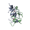


 PDBj
PDBj


 Assembly

 Mass: 18.015 Da / Num. of mol.: 41 / Source method: isolated from a natural source / Formula: H2O
 Sample preparation
 / Beamline: X06DA / Wavelength: 1
  Processing