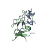
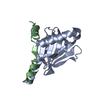
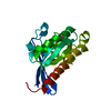
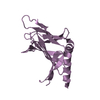
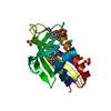
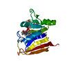
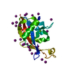
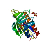
Entry Database : PDB / ID : 3so6Title Crystal structure of the LDL receptor tail in complex with autosomal recessive hypercholesterolemia PTB domain LDL receptor adaptor protein Low-density lipoprotein receptor Keywords / / / / / / / / / Function / homology Function Domain/homology Component
/ / / / / / / / / / / / / / / / / / / / / / / / / / / / / / / / / / / / / / / / / / / / / / / / / / / / / / / / / / / / / / / / / / / / / / / / / / / / / / / / / / / / / / / / / / / / / / / / / / / / / / / / / / / / / / / / / / / / / / / / / / / / / / / / / / / / / / / / / / / / / / / / / / / / / / / / / Biological species Rattus norvegicus (Norway rat)Homo sapiens (human)Method / / / / Resolution : 1.37 Å Authors Dvir, H. / Zajonc, D.M. Journal : Proc.Natl.Acad.Sci.USA / Year : 2012Title : Atomic structure of the autosomal recessive hypercholesterolemia phosphotyrosine-binding domain in complex with the LDL-receptor tail.Authors : Dvir, H. / Shah, M. / Girardi, E. / Guo, L. / Farquhar, M.G. / Zajonc, D.M. History Deposition Jun 29, 2011 Deposition site / Processing site Revision 1.0 Apr 18, 2012 Provider / Type Revision 1.1 May 2, 2012 Group Revision 1.2 May 16, 2012 Group Revision 1.3 Nov 8, 2017 Group / Category / Item Revision 1.4 Feb 28, 2024 Group / Database referencesCategory chem_comp_atom / chem_comp_bond ... chem_comp_atom / chem_comp_bond / database_2 / struct_ref_seq_dif Item / _database_2.pdbx_database_accession / _struct_ref_seq_dif.details
Show all Show less
 Movie
Movie Controller

 Yorodumi
 Open data
 Basic information
 Components
 Keywords
 Function and homology information
 Homo sapiens (human)
 X-RAY DIFFRACTION /
  Authors
 Citation
 Structure visualization
 Downloads & links
 Other downloads
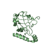
 PDBj
PDBj


 Assembly

 Mass: 18.015 Da / Num. of mol.: 185 / Source method: isolated from a natural source / Formula: H2O
 Sample preparation
 / Beamline: BL11-1 / Wavelength: 0.97945 Å
 Processing