SHEET DETERMINATION METHOD: DSSP THE SHEETS PRESENTED AS "AB" IN EACH CHAIN ON SHEET RECORDS BELOW ... SHEET DETERMINATION METHOD: DSSP THE SHEETS PRESENTED AS "AB" IN EACH CHAIN ON SHEET RECORDS BELOW IS ACTUALLY AN 8-STRANDED BARREL THIS IS REPRESENTED BY A 9-STRANDED SHEET IN WHICH THE FIRST AND LAST STRANDS ARE IDENTICAL.
Protocol: SINGLE WAVELENGTH / Monochromatic (M) / Laue (L): M / Scattering type: x-ray
Radiation wavelength
Wavelength: 0.9795 Å / Relative weight: 1
Reflection
Resolution: 1.6→37.42 Å / Num. obs: 77716 / % possible obs: 100 % / Observed criterion σ(I): 0 / Redundancy: 5.4 % / Biso Wilson estimate: 15.213 Å2 / Rmerge(I) obs: 0.08 / Net I/σ(I): 13.3
Reflection shell
Resolution: 1.6→1.69 Å / Redundancy: 5.4 % / Rmerge(I) obs: 0.44 / Mean I/σ(I) obs: 3.4 / % possible all: 100
-
Processing
Software
Name
Version
Classification
REFMAC
5.5.0109
refinement
MOSFLM
datareduction
SCALA
datascaling
Refinement
Method to determine structure: OTHER Starting model: NONE Resolution: 1.6→37.42 Å / Cor.coef. Fo:Fc: 0.963 / Cor.coef. Fo:Fc free: 0.955 / SU B: 0.962 / SU ML: 0.034 / Cross valid method: THROUGHOUT / ESU R: 0.059 / ESU R Free: 0.059 / Stereochemistry target values: MAXIMUM LIKELIHOOD / Details: HYDROGENS HAVE BEEN ADDED IN THE RIDING POSITIONS.
Rfactor
Num. reflection
% reflection
Selection details
Rfree
0.17633
3897
5 %
RANDOM
Rwork
0.16094
-
-
-
obs
0.16171
73722
99.89 %
-
Solvent computation
Ion probe radii: 0.8 Å / Shrinkage radii: 0.8 Å / VDW probe radii: 1.4 Å / Solvent model: MASK
 Movie
Movie Controller

 Yorodumi
 Open data
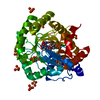
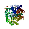
 Basic information
 Components
 Keywords
 Function and homology information
 HOMO SAPIENS (human)
 X-RAY DIFFRACTION /
  Authors
 Citation
 Structure visualization
 Downloads & links
 Other downloads


 PDBj
PDBj
 Assembly
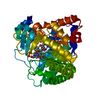


 Mass: 456.344 Da / Num. of mol.: 1 / Source method: obtained synthetically / Formula: C17H21N4O9P
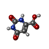
 Mass: 156.096 Da / Num. of mol.: 1 / Source method: obtained synthetically / Formula: C5H4N2O4
 Mass: 96.063 Da / Num. of mol.: 2 / Source method: obtained synthetically / Formula: SO4
 Mass: 35.453 Da / Num. of mol.: 1 / Source method: obtained synthetically / Formula: Cl
 Mass: 341.216 Da / Num. of mol.: 1 / Source method: obtained synthetically / Formula: C13H10Cl2N4OS
 Mass: 59.044 Da / Num. of mol.: 2 / Source method: obtained synthetically / Formula: C2H3O2
 Sample preparation
 / Beamline: I02 / Wavelength: 0.9795
  Processing