 Movie
Movie Controller

[English] 日本語
 Yorodumi
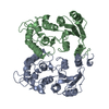
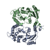
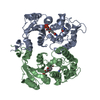
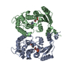
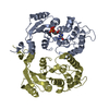
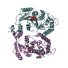
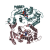
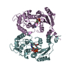
- PDB-3zwn: Crystal structure of Aplysia cyclase complexed with substrate NGD... -
+ Open data

- Basic information
| Entry | Database: PDB / ID: 3zwn | ||||||
|---|---|---|---|---|---|---|---|
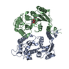
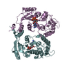
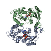
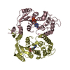
| Title | Crystal structure of Aplysia cyclase complexed with substrate NGD and product cGDPR | ||||||
 Components Components | ADP-RIBOSYL CYCLASE | ||||||
 Keywords Keywords | HYDROLASE | ||||||
| Function / homology |  Function and homology information Function and homology information2'-phospho-ADP-ribosyl cyclase/2'-phospho-cyclic-ADP-ribose transferase / phosphorus-oxygen lyase activity / NAD+ nucleosidase activity, cyclic ADP-ribose generating / Hydrolases; Glycosylases; Hydrolysing N-glycosyl compounds / single fertilization / transferase activity / cytoplasmic vesicle / plasma membrane Similarity search - Function | ||||||
| Biological species |  | ||||||
| Method |  X-RAY DIFFRACTION / SYNCHROTRON / MOLECULAR REPLACEMENT / Resolution: 1.8 Å X-RAY DIFFRACTION / SYNCHROTRON / MOLECULAR REPLACEMENT / Resolution: 1.8 Å | ||||||
 Authors Authors | Kotaka, M. / Graeff, R. / Zhang, L.H. / Lee, H.C. / Hao, Q. | ||||||
 Citation Citation | Journal: J.Mol.Biol. / Year: 2012 Title: Structural Studies of Intermediates Along the Cyclization Pathway of Aplysia Adp-Ribosyl Cyclase. Authors: Kotaka, M. / Graeff, R. / Chen, Z. / Zhang, L.H. / Lee, H.C. / Hao, Q. | ||||||
| History |
|
- Structure visualization
| Structure viewer | Molecule: MolmilJmol/JSmol |
|---|
- Downloads & links
-Download
| PDBx/mmCIF format | 3zwn.cif.gz | 233.9 KB | Display | PDBx/mmCIF format |
|---|---|---|---|---|
| PDB format | pdb3zwn.ent.gz | 186.4 KB | Display | PDB format |
| PDBx/mmJSON format | 3zwn.json.gz | Tree view | PDBx/mmJSON format | |
| Others |  Other downloads Other downloads |
-Validation report
| Arichive directory | https://data.pdbj.org/pub/pdb/validation_reports/zw/3zwnftp://data.pdbj.org/pub/pdb/validation_reports/zw/3zwn | HTTPS FTP |
|---|
-Related structure data
| Related structure data |  3zwmC  3zwoC 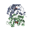 3zwpC 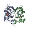 3zwvC 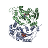 3zwwC 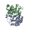 1r12S S: Starting model for refinement C: citing same article ( |
|---|---|
| Similar structure data |


-Links
 PDBj
PDBj- Assembly
| Deposited unit | 
| ||||||||
|---|---|---|---|---|---|---|---|---|---|
| 1 |
| ||||||||
| Unit cell |
| ||||||||
| Noncrystallographic symmetry (NCS) | NCS oper: (Code: given Matrix: (-0.8639, -0.3603, -0.352), Vector: |
-Components
| #1: Protein | Mass: 29722.102 Da / Num. of mol.: 2 Source method: isolated from a genetically manipulated source Source: (gene. exp.) Plasmid: PPICZALPHAA / Production host:  PICHIA PASTORIS (fungus) / Strain (production host): X-33 / References: UniProt: P29241, NAD+ glycohydrolase PICHIA PASTORIS (fungus) / Strain (production host): X-33 / References: UniProt: P29241, NAD+ glycohydrolase#2: Chemical | ChemComp-CGR / | 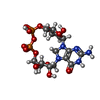  Mass: 559.316 Da / Num. of mol.: 1 / Source method: obtained synthetically / Formula: C15H23N5O14P2 Mass: 559.316 Da / Num. of mol.: 1 / Source method: obtained synthetically / Formula: C15H23N5O14P2#3: Chemical | ChemComp-NGD / |   Mass: 680.432 Da / Num. of mol.: 1 / Source method: obtained synthetically / Formula: C21H28N7O15P2 Mass: 680.432 Da / Num. of mol.: 1 / Source method: obtained synthetically / Formula: C21H28N7O15P2#4: Water | ChemComp-HOH / |  Mass: 18.015 Da / Num. of mol.: 452 / Source method: isolated from a natural source / Formula: H2O Mass: 18.015 Da / Num. of mol.: 452 / Source method: isolated from a natural source / Formula: H2OHas protein modification | Y | Sequence details | THE ADDITIONAL ALANINE AT THE N-TERMINUS ARE ACTUALLY AMINO ACIDS LEFT OVER FROM THE CLEAVAGE OF ...THE ADDITIONAL | |
|---|
-Experimental details
-Experiment
| Experiment | Method: X-RAY DIFFRACTION / Number of used crystals: 1 |
|---|
- Sample preparation
| Crystal | Density Matthews: 2.84 Å3/Da / Density % sol: 56.71 % / Description: NONE |
|---|---|
| Crystal grow | pH: 7.5 / Details: 0.1M IMIDAZOLE, PH 7.5, 12-14% PEG 4000. |
-Data collection
| Diffraction | Mean temperature: 100 K |
|---|---|
| Diffraction source | Source: SYNCHROTRON / Site: NSRRC  / Beamline: BL13B1 / Wavelength: 1 / Beamline: BL13B1 / Wavelength: 1 |
| Detector | Type: ADSC CCD / Detector: CCD / Date: May 19, 2009 |
| Radiation | Protocol: SINGLE WAVELENGTH / Monochromatic (M) / Laue (L): M / Scattering type: x-ray |
| Radiation wavelength | Wavelength: 1 Å / Relative weight: 1 |
| Reflection | Resolution: 1.8→30 Å / Num. obs: 48804 / % possible obs: 94 % / Observed criterion σ(I): 1.4 / Redundancy: 3.4 % / Biso Wilson estimate: 27.2 Å2 / Rmerge(I) obs: 0.08 / Net I/σ(I): 17.5 |
| Reflection shell | Resolution: 1.8→1.86 Å / Rmerge(I) obs: 0.55 / Mean I/σ(I) obs: 1.4 / % possible all: 69.4 |
- Processing
| Software |
| ||||||||||||||||||||||||||||||||||||||||||||||||||||||||||||||||||||||||||||||||||||||||||||||||||||||||||||||||||||||||||||||||||||||||||||||||||||||||||||||||||||||||||||||||||||||
|---|---|---|---|---|---|---|---|---|---|---|---|---|---|---|---|---|---|---|---|---|---|---|---|---|---|---|---|---|---|---|---|---|---|---|---|---|---|---|---|---|---|---|---|---|---|---|---|---|---|---|---|---|---|---|---|---|---|---|---|---|---|---|---|---|---|---|---|---|---|---|---|---|---|---|---|---|---|---|---|---|---|---|---|---|---|---|---|---|---|---|---|---|---|---|---|---|---|---|---|---|---|---|---|---|---|---|---|---|---|---|---|---|---|---|---|---|---|---|---|---|---|---|---|---|---|---|---|---|---|---|---|---|---|---|---|---|---|---|---|---|---|---|---|---|---|---|---|---|---|---|---|---|---|---|---|---|---|---|---|---|---|---|---|---|---|---|---|---|---|---|---|---|---|---|---|---|---|---|---|---|---|---|---|
| Refinement | Method to determine structure: MOLECULAR REPLACEMENT Starting model: PDB ENTRY 1R12 Resolution: 1.8→25 Å / Cor.coef. Fo:Fc: 0.961 / Cor.coef. Fo:Fc free: 0.937 / SU B: 6.003 / SU ML: 0.084 / Cross valid method: THROUGHOUT / ESU R: 0.133 / ESU R Free: 0.137 / Stereochemistry target values: MAXIMUM LIKELIHOOD / Details: HYDROGENS HAVE BEEN ADDED IN THE RIDING POSITIONS.
| ||||||||||||||||||||||||||||||||||||||||||||||||||||||||||||||||||||||||||||||||||||||||||||||||||||||||||||||||||||||||||||||||||||||||||||||||||||||||||||||||||||||||||||||||||||||
| Solvent computation | Ion probe radii: 0.8 Å / Shrinkage radii: 0.8 Å / VDW probe radii: 1.2 Å / Solvent model: MASK | ||||||||||||||||||||||||||||||||||||||||||||||||||||||||||||||||||||||||||||||||||||||||||||||||||||||||||||||||||||||||||||||||||||||||||||||||||||||||||||||||||||||||||||||||||||||
| Displacement parameters | Biso mean: 29.843 Å2
| ||||||||||||||||||||||||||||||||||||||||||||||||||||||||||||||||||||||||||||||||||||||||||||||||||||||||||||||||||||||||||||||||||||||||||||||||||||||||||||||||||||||||||||||||||||||
| Refinement step | Cycle: LAST / Resolution: 1.8→25 Å
| ||||||||||||||||||||||||||||||||||||||||||||||||||||||||||||||||||||||||||||||||||||||||||||||||||||||||||||||||||||||||||||||||||||||||||||||||||||||||||||||||||||||||||||||||||||||
| Refine LS restraints |
|